

Neurocysticercosis
- PMID: 25360206
- PMCID: PMC4212415
- DOI: 10.1177/1941874414533351
Neurocysticercosis
Abstract
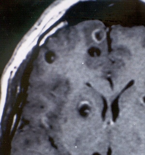
Neurocysticercosis, the most common helminthic infection of the nervous system, is a major cause of acquired epilepsy worldwide. The disease occurs when humans become intermediate hosts of the tapeworm Taenia solium after ingesting its eggs by contagion from an asymptomatic Taenia carrier. Within the nervous system, parasites may locate in brain parenchyma, subarachnoid space, ventricular system, or spinal cord, causing several pathological changes that are responsible for the clinical pleomorphism of the disease. Seizures are the most common clinical manifestation, but a sizable proportion of patients develop focal deficits, intracranial hypertension, or cognitive decline. Preoperative diagnosis of neurocysticercosis is possible after proper integration of data from neuroimaging studies and immunological tests. Cysticidal drugs (albendazole and praziquantel) have changed the prognosis of most patients with neurocysticercosis. The use of these drugs has shown to reduce the parasite load within the central nervous system and to improve the clinical prognosis of the disease in many cases. Future studies should focus on disease eradication through the implementation of control programs against all the interrelated steps in the life cycle of T solium, including human carriers of the adult tapeworm, infected pigs, and eggs in the environment.
Keywords: Taenia solium; cysticercosis; neurocysticercosis.
Conflict of interest statement
Figures


References
-
- Del Brutto OH, Santibañez R, Idrovo L, et al. . Epilepsy and neurocysticercosis in Atahualpa: a door-to-door survey in rural coastal Ecuador. Epilepsia. 2005;46(4):583–587 - PubMed
-
- Medina MT, Duron RM, Martinez L. Prevalence, incidence, and etiology of epilepsies in rural Honduras: the Salama study. Epilepsia. 2005;46(1):124–131 - PubMed
-
- Montano SM, Villaran MV, Ylquimiche L, et al. . Neurocysticercosis. Association between seizures, serology, and brain CT in rural Peru. Neurology. 2005;65(2):229–234 - PubMed
-
- Del Brutto OH, Garcia HH. Neurocysticercosis in nonendemic countries: time for a reappraisal. Neuroepidemiology. 2012;39(2):145–146 - PubMed
-
- Del Brutto OH. A review of cases of human cysticercosis in Canada. Can J Neurol Sci. 2012;39(3):319–322 - PubMed
LinkOut - more resources
Full Text Sources
Other Literature Sources