

Differentiation of ex vivo human breast tissue using polarization-sensitive optical coherence tomography
- PMID: 25360360
- PMCID: PMC4206312
- DOI: 10.1364/BOE.5.003417
Differentiation of ex vivo human breast tissue using polarization-sensitive optical coherence tomography
Abstract
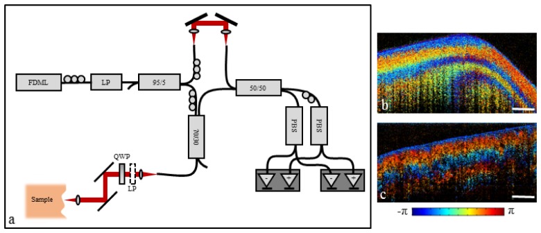
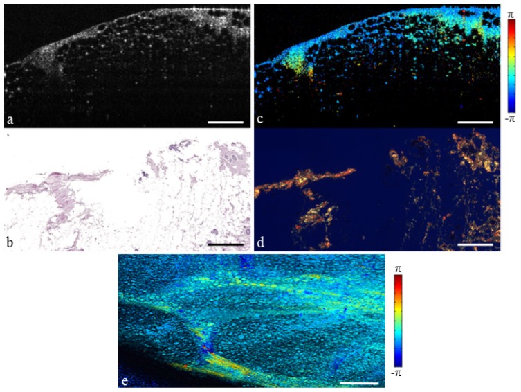
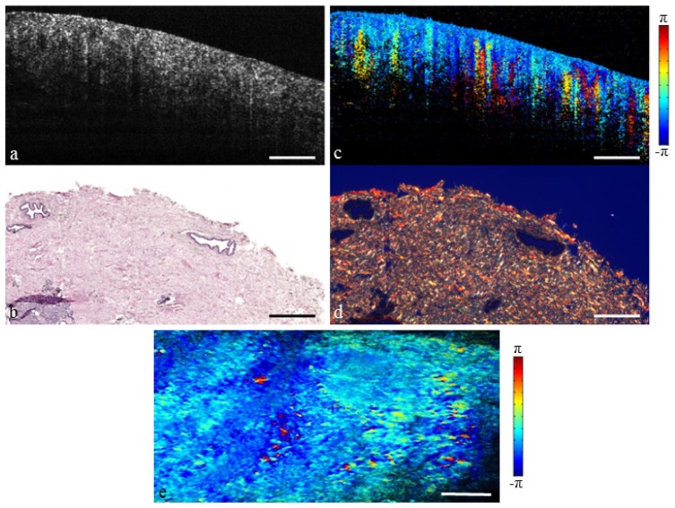
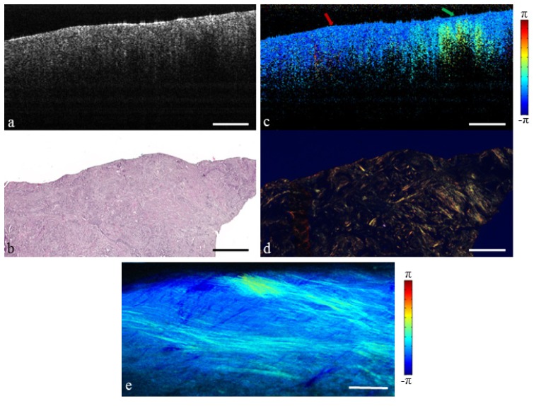
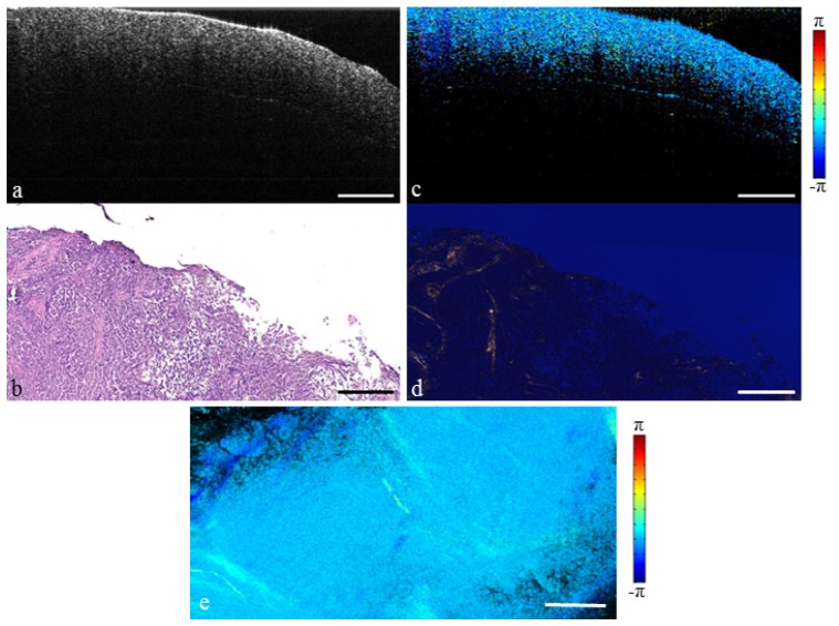
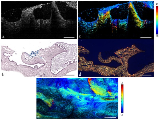
Successful treatment of breast cancer typically requires surgical removal of the tumor. Optical coherence tomography (OCT) has been previously developed for real-time imaging of the surgical margin. However, it can be difficult to distinguish between normal stromal tissue and cancer tissue based on scattering intensity and structure alone. Polarization-sensitive optical coherence tomography (PS-OCT) is sensitive to form birefringence of biological tissue. We report on the development of a high-speed PS-OCT system and imaging of ex vivo human breast tissue, showing enhanced contrast between healthy and cancerous tissues based upon collagen content confirmed with corresponding histology. These results demonstrate the feasibility of using PS-OCT to supplement structural OCT as a possible method for intraoperative tumor margin evaluation.
Keywords: (110.4500) Optical coherence tomography; (110.5405) Polarimetric imaging; (170.3880) Medical and biological imaging; (170.4500) Optical coherence tomography; (170.6935) Tissue characterization; (260.1440) Birefringence.
Figures
Similar articles
-
Determination of characteristics of degenerative joint disease using optical coherence tomography and polarization sensitive optical coherence tomography.Lasers Surg Med. 2006 Oct;38(9):852-65. doi: 10.1002/lsm.20394. Lasers Surg Med. 2006. PMID: 16998913
-
Polarization-sensitive optical coherence tomography for renal tumor detection in ex vivo human kidneys.Opt Lasers Eng. 2024 Feb;173:107900. doi: 10.1016/j.optlaseng.2023.107900. Epub 2023 Oct 23. Opt Lasers Eng. 2024. PMID: 37982078 Free PMC article.
-
Visualization of prostatic nerves by polarization-sensitive optical coherence tomography.Biomed Opt Express. 2016 Aug 1;7(9):3170-3183. doi: 10.1364/BOE.7.003170. eCollection 2016 Sep 1. Biomed Opt Express. 2016. PMID: 27699090 Free PMC article.
-
Polarization sensitive optical coherence tomography - a review [Invited].Biomed Opt Express. 2017 Feb 24;8(3):1838-1873. doi: 10.1364/BOE.8.001838. eCollection 2017 Mar 1. Biomed Opt Express. 2017. PMID: 28663869 Free PMC article. Review.
-
Review of polarization sensitive optical coherence tomography and Stokes vector determination.J Biomed Opt. 2002 Jul;7(3):359-71. doi: 10.1117/1.1483879. J Biomed Opt. 2002. PMID: 12175285 Review.
Cited by
-
Compressive sensing for polarization sensitive optical coherence tomography.J Phys D Appl Phys. 2021 Jul;54(29):294005. doi: 10.1088/1361-6463/abf958. Epub 2021 May 14. J Phys D Appl Phys. 2021. PMID: 38222471 Free PMC article.
-
Polarization-artifact reduction and accuracy improvement of Jones-matrix polarization-sensitive optical coherence tomography by multi-focus-averaging based multiple scattering reduction.Biomed Opt Express. 2023 Dec 18;15(1):256-276. doi: 10.1364/BOE.509763. eCollection 2024 Jan 1. Biomed Opt Express. 2023. PMID: 38223182 Free PMC article.
-
Polarization-sensitive interferometric synthetic aperture microscopy.Appl Phys Lett. 2015 Nov 23;107(21):211106. doi: 10.1063/1.4936236. Appl Phys Lett. 2015. PMID: 26648593 Free PMC article.
-
Review of intraoperative optical coherence tomography: technology and applications [Invited].Biomed Opt Express. 2017 Feb 21;8(3):1607-1637. doi: 10.1364/BOE.8.001607. eCollection 2017 Mar 1. Biomed Opt Express. 2017. PMID: 28663853 Free PMC article.
-
Evaluating optical coherence tomography for surgical margin assessment of canine mammary tumours.Vet Comp Oncol. 2021 Dec;19(4):697-706. doi: 10.1111/vco.12632. Epub 2020 Jul 26. Vet Comp Oncol. 2021. PMID: 32562330 Free PMC article.
References
-
- American Cancer Society , Breast Cancer Facts & Figs. 2013–2014 (American Cancer Society, Atlanta, GA, 2013).
-
- Fisher B., Anderson S., Bryant J., Margolese R. G., Deutsch M., Fisher E. R., Jeong J.-H., Wolmark N., “Twenty-year follow-up of a randomized trial comparing total mastectomy, lumpectomy, and lumpectomy plus irradiation for the treatment of invasive breast cancer,” N. Engl. J. Med. 347(16), 1233–1241 (2002).10.1056/NEJMoa022152 - DOI - PubMed
-
- Veronesi U., Cascinelli N., Mariani L., Greco M., Saccozzi R., Luini A., Aguilar M., Marubini E., “Twenty-year follow-up of a randomized study comparing breast-conserving surgery with radical mastectomy for early breast cancer,” N. Engl. J. Med. 347(16), 1227–1232 (2002).10.1056/NEJMoa020989 - DOI - PubMed
Grants and funding
LinkOut - more resources
Full Text Sources
Other Literature Sources