. 2014 Sep 4;5(10):3427-33.
doi: 10.1364/BOE.5.003427. eCollection 2014 Oct 1.

Measurements of multiphoton action cross sections for multiphoton microscopy

Affiliations

Measurements of multiphoton action cross sections for multiphoton microscopy

Li-Chung Cheng et al. Biomed Opt Express. .

Abstract

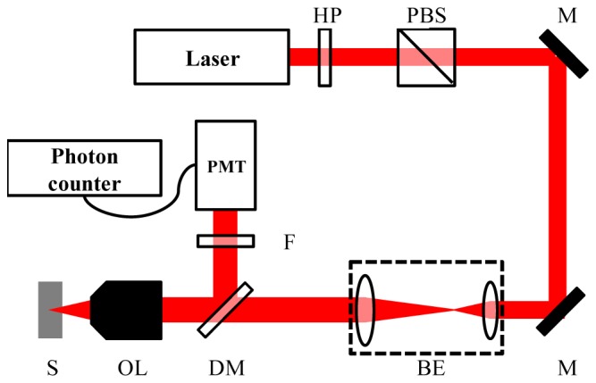
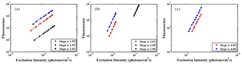
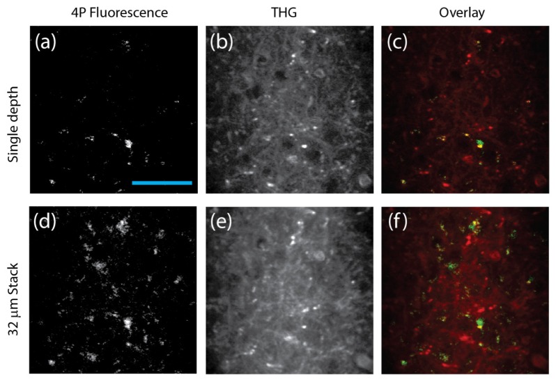
We report quantitative measurements of two-, three-, and four-photon excitation action cross sections of several commonly used fluorophores and fluorescent proteins at three different excitation wavelengths of 800 nm, 1300 nm, and 1680 nm. The measured cross section values are consistent with simple quantum mechanic estimations. These values indicate that the optimum repetition rate for deep tissue 3-photon microscopy is approximately 1 to 2 MHz. We further demonstrate that it is feasible to perform 4-photon fluorescence microscopy of GFP labeled microglia in mouse brain in vivo at 1700 nm. 4-photon excitation increases the accessibility of fluorophores at the long wavelength spectral window of 1700 nm.

Keywords: (190.0190) Nonlinear optics; (190.4180) Multiphoton processes.


Figures

Fig. 1
Fig. 1
Experiment setup for measuring two-, three-, and four-photon excitation cross section. HP: half-wave plate. PBS: polarization beam splitter. M: mirror. BE: beam expander. DM: dichroic mirror. OL: objective lens. S: sample. F: emission filter. PMT: photomultiplier tube.
Fig. 2
Fig. 2
Logarithmic plots of the dependence of (a) two-, (b) three-, and (c) four-photon-excited fluorescence on excitation intensity for fluorescein (red square), wtGFP (blue square), and SR101 (black square). The slopes are indicated in the lower-right corner of each figure.
Fig. 3
Fig. 3
4PM and THG images of a mouse brain in vivo. (a-c): 2-frame averaging at a depth of 472 μm below the surface of the brain. (d-f): Average intensity of a 32 μm stack (2 frames per depth, 4 μm step size) ranging from 456 to 484 μm below the surface of the brain. The acquisition time was 4 seconds per frame, and the average power was 23 mW (repetition rate 1.3 MHz) on the brain surface. The scale bar is 50 μm.

References

    1. Kobat D., Durst M. E., Nishimura N., Wong A. W., Schaffer C. B., Xu C., “Deep tissue multiphoton microscopy using longer wavelength excitation,” Opt. Express 17(16), 13354–13364 (2009).10.1364/OE.17.013354 - DOI - PubMed
    1. D. Kobat, N.G. Horton, and C. Xu, “In vivo two-photon imaging of cortical vasculature in mice to 1.5-mm depth with 1280-nm excitation,” CLEO, postdeadline paper, PDPB3(2011).
    1. Balu M., Baldacchini T., Carter J., Krasieva T. B., Zadoyan R., Tromberg B. J., “Effect of excitation wavelength on penetration depth in nonlinear optical microscopy of turbid media,” J. Biomed. Opt. 14(1), 010508 (2009).10.1117/1.3081544 - DOI - PMC - PubMed
    1. Andresen V., Alexander S., Heupel W. M., Hirschberg M., Hoffman R. M., Friedl P., “Infrared multiphoton microscopy: subcellular-resolved deep tissue imaging,” Curr. Opin. Biotechnol. 20(1), 54–62 (2009).10.1016/j.copbio.2009.02.008 - DOI - PubMed
    1. Kobat D., Horton N. G., Xu C., “In vivo two-photon microscopy to 1.6-mm depth in mouse cortex,” J. Biomed. Opt. 16(10), 106014 (2011).10.1117/1.3646209 - DOI - PubMed

LinkOut - more resources