. 2014 Oct 31;9(10):e111632.
doi: 10.1371/journal.pone.0111632. eCollection 2014.

LRRK2 transport is regulated by its novel interacting partner Rab32

Affiliations

LRRK2 transport is regulated by its novel interacting partner Rab32

Dieter Waschbüsch et al. PLoS One. .

Abstract

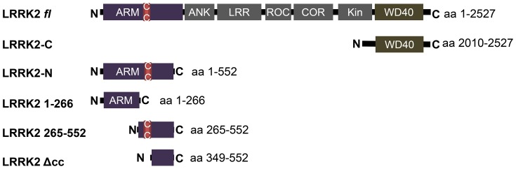
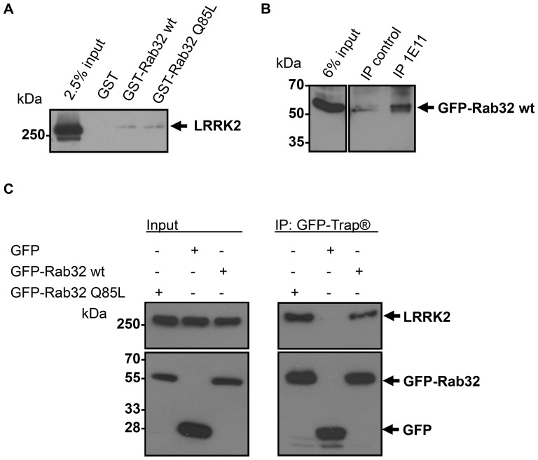
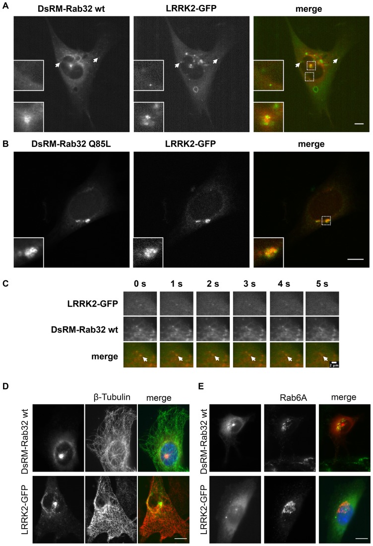
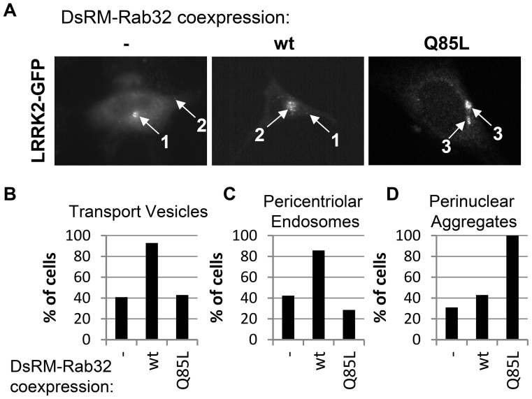
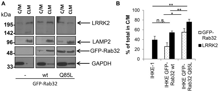
Leucine-rich repeat kinase 2 (LRRK2) is a multi-domain 280 kDa protein that is linked to Parkinson's disease (PD). Mutations especially in the GTPase and kinase domains of LRRK2 are the most common causes of heritable PD and are also found in sporadic forms of PD. Although the cellular function of LRRK2 is largely unknown there is increasing evidence that these mutations cause cell death due to autophagic dysfunction and mitochondrial damage. Here, we demonstrate a novel mechanism of LRRK2 binding and transport, which involves the small GTPases Rab32 and Rab38. Rab32 and its closest homologue Rab38 are known to organize the trans-Golgi network and transport of key enzymes in melanogenesis, whereas their function in non-melanogenic cells is still not well understood. Cellular processes such as autophagy, mitochondrial dynamics, phagocytosis or inflammatory processes in the brain have previously been linked to Rab32. Here, we demonstrate that Rab32 and Rab38, but no other GTPase tested, directly interact with LRRK2. GFP-Trap analyses confirmed the interaction of Rab32 with the endogenous LRRK2. In yeast two-hybrid experiments we identified a predicted coiled-coil motif containing region within the aminoterminus of LRRK2 as the possible interacting domain. Fluorescence microscopy demonstrated a co-localization of Rab32 and LRRK2 at recycling endosomes and transport vesicles, while overexpression of a constitutively active mutant of Rab32 led to an increased co-localization with Rab7/9 positive perinuclear late endosomes/MVBs. Subcellular fractionation experiments supported the novel role of Rab32 in LRRK2 late endosomal transport and sorting in the cell. Thus, Rab32 may regulate the physiological functions of LRRK2.


Conflict of interest statement

Competing Interests: The authors declare that they have no competing interests.

Figures

Figure 1
Figure 1. LRRK2 constructs used in this study.
ARM: armadillo repeats; ANK: ankyrin repeats; LRR: leucine rich repeats; ROC: Ras of complex; COR: C-terminal of ROC; Kin: kinase domain; WD40: WD40 domain; c/c: coiled coil motif (aa 319–348).
Figure 2
Figure 2. Binding of endogenous LRRK2 by the small GTPase Rab32.
(A) GST-Rab32 wt, GST-Rab32 Q85L or GST as control was applied to glutathione agarose beads followed by incubation with NIH3T3 lysate overnight. Samples were analyzed by 6% SDS-PAGE and subsequent Western blot analysis to detect LRRK2. n≥3 independent experiments. (B) Lysates from IHKE-1 cells stably expressing GFP-Rab32 wt were incubated overnight with an anti-LRRK2 antibody (1E11). IP control = no antibody was added. Co-precipitated GFP-Rab32 wt was detected using an anti-Rab32 antibody. n = 3 independent experiments. (C) Lysates from IHKE-1 cells expressing GFP-Rab32 wt, GFP-Rab32 Q85L or GFP as control were subjected to immunoprecipitation by the GFP-Trap kit. Co-precipitated endogenous LRRK2 was detected using an anti LRRK2 antibody. n = 2 independent experiments.
Figure 3
Figure 3. Co-localization analysis of LRRK2-GFP and DsRed-Monomer-Rab32.
(A) NIH3T3 cells were co-transfected with plasmids encoding DsRed-Monomer-Rab32 wt and LRRK2-GFP. Living cells were imaged using a Zeiss LSM5 live microscope. The image shows a still frame from Movie S1. Scale bar = 10 µm. (B) NIH3T3 cells were co-transfected with plasmids encoding DsRed-Monomer-Rab32 Q85L and LRRK2-GFP. After 48 hours the cells were fixed in 4% PFA and subsequently analyzed with a laser scanning microscope. Scale bar = 10 µm. (C) Co-transport and sorting of LRRK2-GFP by DsRed-Monomer-Rab32 wt. Image series are a detail view from Movie S2. Both channels were recorded simultaneously every second. Scale bar = 2 µm. (D and E) NIH3T3 cells expressing either DsRed-Monomer-Rab32 wt or LRRK2-GFP were fixed and subsequently subjected to immunofluorescence labeling of β-tubulin (D) or the Golgi marker Rab6A (E). Scale bar = 10 µm.
Figure 4
Figure 4. Co-localization analysis of Rab32 wt and the constitutively active mutant Rab32 Q85L with different endosomal markers.
(A) NIH3T3 cells were co-transfected with plasmids encoding the recycling endosome marker GFP-Rab11B and DsRed-Monomer-Rab32 wt or DsRed-Monomer-Rab32 Q85L, fixed and analyzed by fluorescence microscopy. Scale bar = 10 µm. (B) Cells were transfected with plasmids encoding for GFP-Rab32 wt or GFP-Rab32 Q85L followed by fixation and subsequent immunofluorescence staining of Rab7. Scale bar = 10 µm. (C–E) NIH3T3 cells expressing either GFP-Rab32 wt or GFP-Rab32 Q85L were fixed and stained for Rab7. (C) Microscopic analysis of GFP-Rab32 Q85L that co-localized with endogenous Rab7 (arrows) in the perinuclear area. Non co-localizing Rab7 was indicated by arrowheads. The perinuclear area was defined by the red (outlines nucleus) and the yellow line (outer border for perinuclear area). The image illustrates the cellular area used for the following analysis. (D) Rab7 perinuclear aggregates co-localizing with GFP-Rab32 wt or GFP-Rab32 Q85L and non co-localizing ones. ctrl. = control (untransfected IHKE-1 cells) (E) Quantification of perinuclear Rab7-positive structures. ctrl. (control): perinuclear Rab7 in untransfected cells; wt/Q85L: perinuclear Rab7 co-localizing with GFP-Rab32 constructs. control/Rab32 wt/Rab32 Q85L: n = 1660/18/167 structures in 67/18/60 cells, 1/1/3 independent experiments. Statistical significance was tested by a Student's T-test: pcontrol-wt = 0.02; pcontrol-Q85L<0.005; pwt-Q85L = 0.02. Scale bar = 10 µm.
Figure 5
Figure 5. LRRK2-GFP subcellular localization in DsRed-Monomer-Rab32 wt and DsRed-Monomer-Rab32 Q85L overexpressing NIH3T3 cells.
(A) Graphical representation of the most common subcellular features of LRRK2-GFP in DsRed-Monomer-Rab32 wt or DsRed-Monomer-Rab32 Q85L expressing cells. 1: pericentriolar endosome; 2: transport vesicles; 3: perinuclear aggregates. (B–D) Microscopic analysis of NIH3T3 cells co-transfected with plasmids encoding DsRed-Monomer-Rab32 wt or DsRed-Monomer-Rab32 Q85L and LRRK2-GFP. In every cell the LRRK2-GFP channel was analyzed for the occurrence of the features transport vesicles, pericentriolar endosomes and perinuclear aggregates. (-) LRRK2-GFP alone: n = 71 cells from 5 independent experiments; LRRK2-GFP and DsRed-Monomer-Rab32 wildtype: n = 15 cells from 3 independent experiments, LRRK2-GFP and DsRed-Monomer-Rab32 Q85L: n = 21 cells from 5 independent experiments.
Figure 6
Figure 6. Subcellular fractionation analysis of endogenous LRRK2 in IHKE-1 cells overexpressing GFP-Rab32 wt or GFP-Rab32 Q85L.
(A) IHKE-1 cells either untransfected (-) or stably overexpressing GFP-Rab32 wt or GFP-Rab32 Q85L were mechanically disrupted and fractionated by differential centrifugation. Fractions received either contained lysosomes and mitochondria (CLM) or cytosol and light membranes (C/M). Western blot analysis to detect the lysosomal marker LAMP2, GAPDH, Rab32 and LRRK2 were performed with the fractions derived from the different IHKE-1 cells. (B) Quantification of Western blot signal intensities using the gel analyzer functionality of ImageJ. The signal intensity for both the C/M and the CLM fraction was added and set to 100%. The graph shows the % of GFP-Rab32 and endogenous LRRK2 in the C/M fraction of the different cell lines. n = 6 independent experiments. Error bars represent S.E.M.; n.s. = not significant; * = p<0.05; ** = p<0.01.

References

    1. West AB, Moore DJ, Biskup S, Bugayenko A, Smith WW, et al. (2005) Parkinson's disease-associated mutations in leucine-rich repeat kinase 2 augment kinase activity. Proc Natl Acad Sci U S A 102: 16842–16847. - PMC - PubMed
    1. Webber PJ, West AB (2009) LRRK2 in Parkinson's disease: function in cells and neurodegeneration. FEBS J 276: 6436–6444. - PMC - PubMed
    1. Gloeckner CJ, Kinkl N, Schumacher A, Braun RJ, O'Neill E, et al. (2006) The Parkinson disease causing LRRK2 mutation I2020T is associated with increased kinase activity. Hum Mol Genet 15: 223–232. - PubMed
    1. Santpere G, Ferrer I (2009) LRRK2 and neurodegeneration. Acta Neuropathol 117: 227–246. - PubMed
    1. Plowey ED, Cherra SJ 3rd, Liu YJ, Chu CT (2008) Role of autophagy in G2019S-LRRK2-associated neurite shortening in differentiated SH-SY5Y cells. J Neurochem 105: 1048–1056. - PMC - PubMed

Publication types

MeSH terms