. 2014 Oct 31;9(10):e109728.
doi: 10.1371/journal.pone.0109728. eCollection 2014.

Establishment of a rabbit Oct4 promoter-based EGFP reporter system

Affiliations

Establishment of a rabbit Oct4 promoter-based EGFP reporter system

Longquan Quan et al. PLoS One. .

Abstract

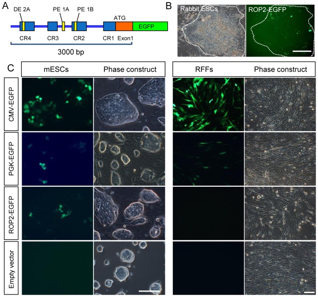
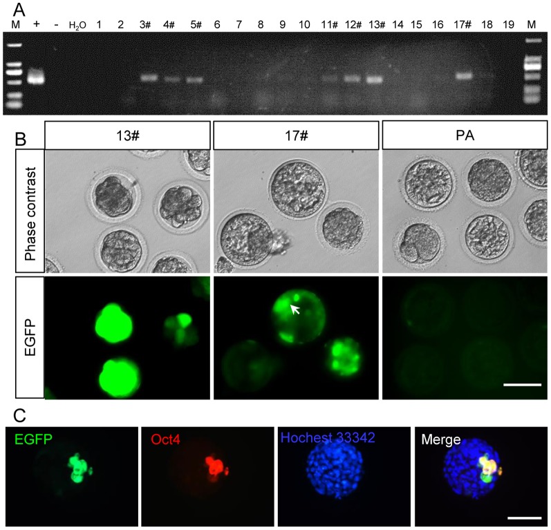
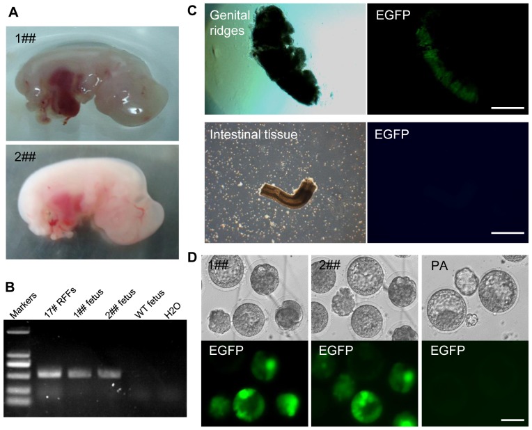
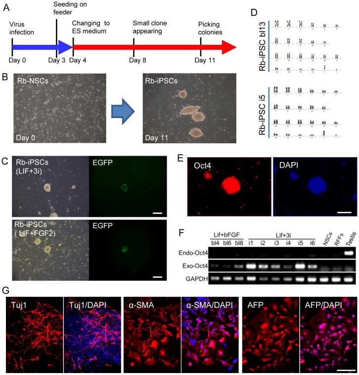
Rabbits are commonly used as laboratory animal models to investigate human diseases and phylogenetic development. However, pluripotent stem cells that contribute to germline transmission have yet to be established in rabbits. The transcription factor Oct4, also known as Pou5f1, is considered essential for the maintenance of the pluripotency of stem cells. Hence, pluripotent cells can be identified by monitoring Oct4 expression using a well-established Oct4 promoter-based reporter system. This study developed a rabbit Oct4 promoter-based enhanced green fluorescent protein (EGFP) reporter system by transfecting pROP2-EGFP into rabbit fetal fibroblasts (RFFs). The transgenic RFFs were used as donor cells for somatic cell nuclear transfer (SCNT). The EGFP expression was detected in the blastocysts and genital ridges of SCNT fetuses. Fibroblasts and neural stem cells (NSCs) were derived from the SCNT fetuses. EGFP was also reactivated in blastocysts after the second SCNT, and induced pluripotent stem cells (iPSCs) were obtained after reprogramming using Yamanaka's factors. The results above indicated that a rabbit reporter system used to monitor the differentiating status of cells was successfully developed.


Conflict of interest statement

Competing Interests: The authors have declared that no competing interests exist.

Figures

Figure 1
Figure 1. Construction of a rabbit Oct4 promoter-based EGFP vector.
(A) Diagram of the pROP2-EGFP vector containing a 3.0 kb rabbit Oct4 promoter followed by EGFP in our experiments. (B) Partial EGFP expression in Rb-ESCs after transfection with pROP2-EGFP. (C) EGFP images after the transfection of CMV-GFP, PGK-EGFP, pROP2-EGFP and empty vector into murine ES cell line (R1) and rabbit fibroblasts (RFFs). Scale bars = 50 µm.
Figure 2
Figure 2. Selection of RFF donor cells stably transfected with pROP2-EGFP for nuclear transfer.
(A) PCR analysis of rabbit fibroblasts stably transfected with pROP2-EGFP. (+: positive control; –: negative control; 1–19: G418-resistant clones; 3#, 4#, 5#, 11#, 12#, 13# and 17#: positive transgenic clones). (B) EGFP expressed in NT embryos using 13#, 17# fibroblasts as nuclear donors, while parthenogenetic blastocysts did not express EGFP. The arrow points ICM. PA: parthenogenetic embryos. (C) Immunoflorescent assay showed that EGFP co-expressed with Oct4 in the ICM from NT embryos at the late blastocyst stage. Scale bars = 100 µm.
Figure 3
Figure 3. Analysis of NT fetuses.
(A) Two cloned fetuses (named 1## and 2##) derived from 17# transgenic clones by SCNT at 15 days after transplantation. (B) Genomic PCR analysis of fetuses 1## and 2## stably transfected with pROP2-EGFP. NC: negative control. (C) EGFP expressed in genital ridges isolated from 2## fetus but not in other tissues, such as intestinal tissues. Scale bars = 1 mm. (D) EGFP reactivated in morulae and blastocysts after the second SCNT was performed using Oct4-EGFP transgenic fibroblasts isolated from fetus 1## and 2## as donors. Parthenogenetic blastocysts did not express EGFP. Scale bars = 100 µm.
Figure 4
Figure 4. Generation of Rb-iPSCs from transgenic Rb-NSCs.
(A) Experimental outline for generating Rb-iPSCs from Rb-NSCs. (B) The morphology of NSCs changed to multi-layer compacted clones after 11 days of induction using human transcription factors Oct4, Klf4, Sox2 and c-Myc. (C) EGFP activated in several Rb-iPS colonies in 3i medium (hLIF plus 3i) and BL medium (hLIF plus bFGF). (D) Rb-iPSCs (Rb-iPSC bl13 cultured in BL medium and Rb-iPSC i5 cultured in 3i medium) maintained a normal karyotype at 15 passages. (E) Immunostaining assay showed the expressed Oct4 in Rb-iPSCs. (F) RT-PCR result demonstrating that endogenous Oct4 was not expressed in all the proliferated colonies and that exogenous Oct4 was continuously expressed in Rb-iPSC colonies. (G) Expressed markers of the three germ layers after Rb-iPSCs differentiated in vitro. Tuj1 (ectoderm); AFP (endoderm); and smooth muscle actin (mesoderm). Scale bars = 50 µm.

References

    1. Ezashi T, Telugu BPVL, Alexenko AP, Sachdev S, Sinha S, et al. (2009) Derivation of induced pluripotent stem cells from pig somatic cells. Proceedings of the National Academy of Sciences of the United States of America 106: 10993–10998. - PMC - PubMed
    1. Honda A, Hirose M, Hatori M, Matoba S, Miyoshi H, et al. (2010) Generation of induced pluripotent stem cells in rabbits: potential experimental models for human regenerative medicine. Journal of Biological Chemistry 285: 31362–31369. - PMC - PubMed
    1. Liao J, Cui C, Chen S, Ren J, Chen J, et al. (2009) Generation of induced pluripotent stem cell lines from adult rat cells. Cell Stem Cell 4: 11–15. - PubMed
    1. Takahashi K, Tanabe K, Ohnuki M, Narita M, Ichisaka T, et al. (2007) Induction of pluripotent stem cells from adult human fibroblasts by defined factors. Cell 131: 861–872. - PubMed
    1. Takahashi K, Yamanaka S (2006) Induction of Pluripotent Stem Cells from Mouse Embryonic and Adult Fibroblast Cultures by Defined Factors. Cell 126: 663–676. - PubMed

Publication types

MeSH terms

Substances