. 2014 Oct 31;9(10):e111496.
doi: 10.1371/journal.pone.0111496. eCollection 2014.

Enterovirus-71 virus-like particles induce the activation and maturation of human monocyte-derived dendritic cells through TLR4 signaling

Affiliations

Enterovirus-71 virus-like particles induce the activation and maturation of human monocyte-derived dendritic cells through TLR4 signaling

Yu-Li Lin et al. PLoS One. .

Abstract

Enterovirus 71 (EV71) causes seasonal epidemics of hand-foot-and-mouth disease and has a high mortality rate among young children. We recently demonstrated potent induction of the humoral and cell-mediated immune response in monkeys immunized with EV71 virus-like particles (VLPs), with a morphology resembling that of infectious EV71 virions but not containing a viral genome, which could potentially be safe as a vaccine for EV71. To elucidate the mechanisms through which EV71 VLPs induce cell-mediated immunity, we studied the immunomodulatory effects of EV71 VLPs on human monocyte-derived dendritic cells (DCs), which bind to and incorporate EV71 VLPs. DC treatment with EV71 VLPs enhanced the expression of CD80, CD86, CD83, CD40, CD54, and HLA-DR on the cell surface; increased the production of interleukin (IL)-12 p40, IL-12 p70, and IL-10 by DCs; and suppressed the capacity of DCs for endocytosis. Treatment with EV71 VLPs also enhanced the ability of DCs to stimulate naïve T cells and induced secretion of interferon (IFN)-γ by T cells and Th1 cell responses. Neutralization with antibodies against Toll-like receptor (TLR) 4 suppressed the capacity of EV71 VLPs to induce the production of IL-12 p40, IL-12 p70, and IL-10 by DCs and inhibited EV71 VLPs binding to DCs. Our study findings clarified the important role for TLR4 signaling in DCs in response to EV71 VLPs and showed that EV71 VLPs induced inhibitor of kappaB alpha (IκBα) degradation and nuclear factor of kappaB (NF-κB) activation.


Conflict of interest statement

Competing Interests: The authors have declared that no competing interests exist.

Figures

Figure 1
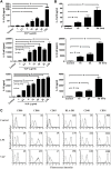
Figure 1. Binding of EV71 VLPs to immature human monocyte-derived DCs.
For the analysis of concentration-dependent effects, human monocyte-derived DCs were incubated with 1 or 10µg/mL EV71 VLPs for 30min on ice to allow binding to the cell surface. For the analysis of time-dependent effects, DCs were incubated with 5µg/mL of EV71 VLPs for 30 or 120min on ice. Detection was facilitated using an anti-EV71 Ab and anti-mouse-IgG Ab (labeled with Alexa Fluor 488). Levels of fluorescence were determined using immunofluorescence confocal microscopy (A) and FACS analysis (B, C). (B) and (C) indicate the respective concentration-dependent and time-dependent effects of the binding of EV71 VLPs to immature human monocyte-derived DCs. The control cells (no VLPs; filled histogram) were stained with the same primary antibody (anti-EV71 Ab) followed by a secondary antibody. (D) and (E) indicate the bar diagram of mean fluorescence intensity (MFI) with statistics for concentration- and time-dependent binding of EV71 VLPs to DCs. Representative data from three different donors with similar results are shown. Values represent the means ± SEs of triplicate samples, and asterisks indicate statistically significant differences (*, P<0.05).
Figure 2
Figure 2. Uptake of EV71 VLPs by immature human monocyte-derived DCs.
For the analysis of concentration-dependent effects, DCs were collected and incubated with different concentrations of CFDA-labeled EV71 VLPs (0, 2, 5, 10, or 20µg/mL) for 120min at 37°C. For the analysis of time-dependent effects, DCs were incubated with 10µg/mL of CFDA-labeled EV71 VLPs for different time points (0, 15, 30, or 120min) at 37°C. At the end of the incubation time, fractions were collected and fixed before double staining with an allophycocyanin-labeled anti-CD1a Ab. Fluorescence was examined by immunofluorescence confocal microscopy (an enlargement of a single cell is shown) (A) and FACS analysis (B, C). (B) and (C) indicate the respective concentration-dependent and time-dependent uptake of EV71 VLPs to immature human monocyte-derived DCs. (D) and (E) show bar diagrams of MFIs with statistics for concentration- and time-dependent uptake of EV71 VLPs. Representative data from three different donors with similar results are shown. Values represent the means ± SEs of triplicate samples, and asterisks indicate statistically significant differences (*, P<0.05).
Figure 3
Figure 3. EV71 VLPs increased cytokines production and enhanced the abundance of cell-surface molecules in human DCs.
(A) Human DCs were cultured for 24h in the presence of 100ng/mL LPS or various concentrations of EV71 VLPs. At the end of the incubation time, the culture medium was collected, and IL-12 p70, IL-12 p40, and IL-10 levels were measured using ELISA. (B) Human DCs were incubated with EV71 VLPs (10µg/mL) for 16, 24, or 48h. At the end of the incubation time, levels of secreted IL-12 p70, IL-12 p40, and IL-10 were analyzed by ELISA. (C) Human DCs were treated with EV71 VLPs (10µg/mL; VLP), LPS (100ng/mL; LPS), or medium alone (Control) for 24h, and levels of surface markers were analyzed by flow cytometry (dotted line, isotype control; solid line, specific mAbs). The values shown in the flow cytometry profiles are the mean fluorescence intensity indices. Representative data from three different donors with similar results are shown. Values represent the means ± SEs of triplicate samples, and asterisks indicate statistically significant differences (*, P<0.05).
Figure 4
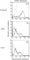
Figure 4. Effects of EV71 VLPs on the endocytic capacity of human monocyte-derived DCs.
At day 5, immature DCs were stimulated with medium alone (Control), LPS (100ng/mL; LPS), or EV71 VLPs (10µg/mL; VLP) for 24h and then incubated with FITC-dextran for 1h at 4°C (dotted lines) or 37°C (solid lines). This experiment was repeated three times (from three different donors) with similar results. The values shown in the flow cytometry profiles are the mean fluorescence intensity indices.
Figure 5
Figure 5. EV71 VLPs enhanced T-cell responses.
(A) Immature DCs were stimulated with medium alone (Control), LPS (100ng/mL), or EV71 VLPs (10 and 20µg/mL; VLP) for 24h. Proliferation of autologous T cells was measured after 5 days of coculture with DCs. (B) Supernatants were analyzed for IFN-γ and IL-5 produced by activated T cells after 2 days of culture. Representative data from three different donors with similar results are shown. Values represent the means ± SEs of triplicate samples, and asterisks indicate statistically significant differences (*, P<0.05).
Figure 6
Figure 6. TLR4 mediated the maturation of DCs induced by EV71 VLPs.
(A) Human DCs were pre-incubated with 20µg/mL of anti-TLR2, anti-TLR4, or IgG2a (as an isotype control) separately for 1h and were then challenged with EV71 VLPs (10µg/mL) for 16h. The control was not treated with EV71 VLPs. Supernatants from the cell cultures were collected to determine levels of IL-12 p70, IL-12 p40, and IL-10. Significant differences in the levels of cytokines produced by DCs treated with Abs and those treated with IgG2a are indicated by asterisks (*p<0.05). (B) Human DCs were pre-incubated with 20µg/mL of anti-TLR4 mAb or IgG2a (as an isotype control) separately for 1h and were then treated with 10µg/mL EV71 VLPs for 30min on ice to allow binding to the cell surface. Surface-bound EV71 VLPs were measured with flow cytometry through immunostaining with anti-EV71 monoclonal antibodies. The control cells (no VLPs; dotted line) were stained with the same primary antibody followed by a secondary antibody. The bar diagram shows the MFI with statistics for the anti-TLR4 mAb effect on binding of EV71 VLPs to DCs. (C) HEK293 cells were transfected with control plasmids, TLR4, or TLR4+MD2. The transfected cells were stimulated with medium alone, LPS (50ng/mL), or EV71 VLP (10µg/mL) for 24h, and IL-8 protein in the supernatants was measured by ELISA. This experiment was repeated three times with similar results. (D) Proteinase K-treated EV71 VLPs abrogated the production of IL-12 p70, IL-12 p40, and IL-10 by DCs. Human DCs were incubated with medium alone (Control), proteinase K (10ng/mL), EV71 VLPs (10µg/mL), proteinase K-treated EV71 VLPs, LPS (100ng/mL), or proteinase K-treated LPS for 24h. At the end of the incubation time, the production of IL-12 p70, IL-12 p40, and IL-10 was analyzed by ELISA. Representative data from three different donors with similar results are shown. Values represent the means ± SEs of triplicate samples, and asterisks indicate statistically significant differences (*, P<0.05).
Figure 7
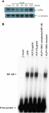
Figure 7. Induction of IκBα degradation and NF-κB activation by incubation of human DCs with EV71 VLPs.
(A) Human DCs were treated with EV71 VLPs (10µg/mL) for the indicated times. Western blotting was used to analyze cytosolic fractions for degradation of IκBα. The lower panel shows anti-α-tubulin staining of the same blot and confirmed equal loading of all samples. (B) Human monocyte-derived DCs were not treated (Cont) or were treated with LPS (100ng/mL) or EV71 VLPs (5 or 10µg/mL) for 2h, and nuclear fractions were prepared. An electrophoretic mobility shift assay was used to assay NF-κB binding activity in the nuclear fractions. To assess the specificity of the binding, a 100-fold excess of cold NF-κB probe (a double-stranded NF-κB oligonucleotide, 5′-AGTTGAGGGGACTTTCCCAGGC-3′) or mutant probe (a double-stranded mutated oligonucleotide, 5′-AGTTGAGGCGACTTTCCCAGGC-3′) was added to the EV71 VLP condition. A double-stranded mutated probe, 5′-AGTTGAGGCGACTTTCCCAGGC-3′, was used to examine the specificity of binding of NF-κB to DNA (the underlined sequence is identical to the κB consensus sequence except for a G-to-C substitution in the NF-κB DNA binding motif). This experiment was repeated three times (from three different donors) with similar results.
Figure 8
Figure 8. The maturation and activation of DCs induced by EV71 VLPs required TLR4 and NF-κB activity and enabled naïve T cells to initiate Th1-cell responses.

References

    1. Tagaya I, Takayama R, Hagiwara A (1981) A large-scale epidemic of hand, foot and mouth disease associated with enterovirus 71 infection in Japan in 1978. Jpn J Med Sci Biol 34: 191–196. - PubMed
    1. Wang SM, Lei HY, Yu CK, Wang JR, Su IJ, et al. (2008) Acute chemokine response in the blood and cerebrospinal fluid of children with enterovirus 71-associated brainstem encephalitis. J Infect Dis 198: 1002–1006. - PubMed
    1. Wu WH, Kuo TC, Lin YT, Huang SW, Liu HF, et al. (2013) Molecular epidemiology of enterovirus 71 infection in the central region of Taiwan from 2002 to 2012. PLoS One 8: e83711. - PMC - PubMed
    1. Wu CN, Lin YC, Fann C, Liao NS, Shih SR, et al. (2001) Protection against lethal enterovirus 71 infection in newborn mice by passive immunization with subunit VP1 vaccines and inactivated virus. Vaccine 20: 895–904. - PubMed
    1. McMinn PC (2002) An overview of the evolution of enterovirus 71 and its clinical and public health significance. FEMS Microbiol Rev 26: 91–107. - PubMed

Publication types