. 2014 Oct 31;9(10):e111575.
doi: 10.1371/journal.pone.0111575. eCollection 2014.

Lichen secondary metabolites in Flavocetraria cucullata exhibit anti-cancer effects on human cancer cells through the induction of apoptosis and suppression of tumorigenic potentials

Affiliations

Lichen secondary metabolites in Flavocetraria cucullata exhibit anti-cancer effects on human cancer cells through the induction of apoptosis and suppression of tumorigenic potentials

Thanh Thi Nguyen et al. PLoS One. .

Expression of concern in

Abstract

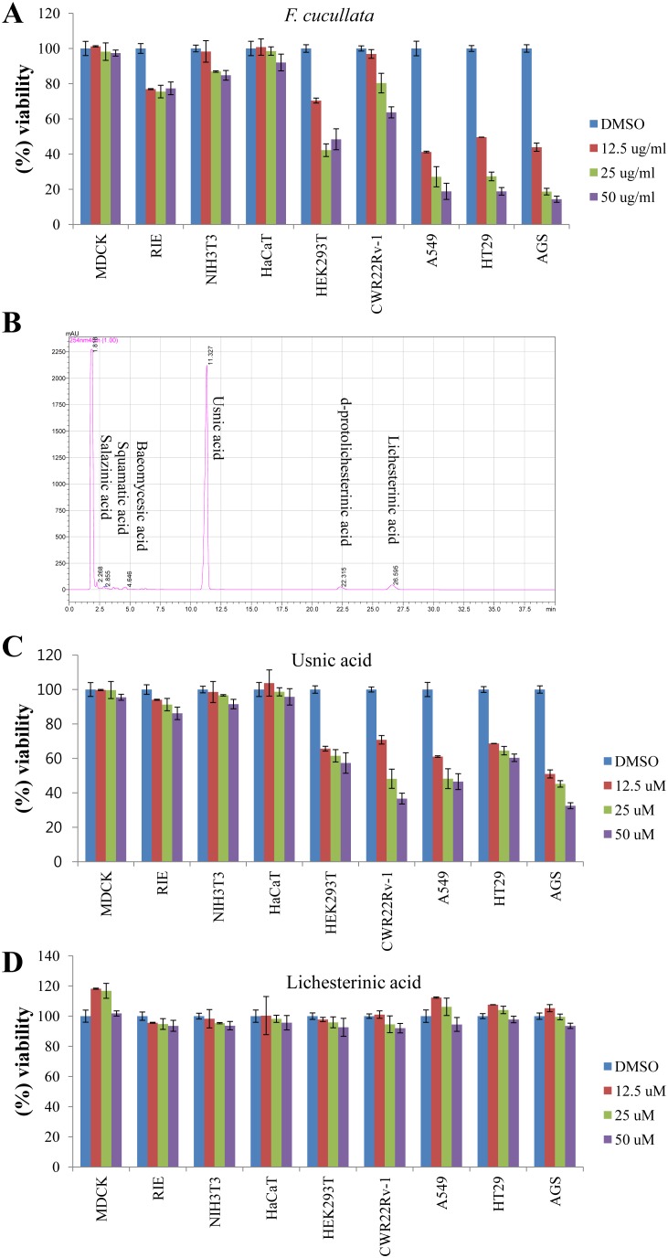
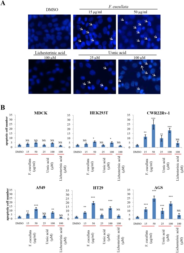
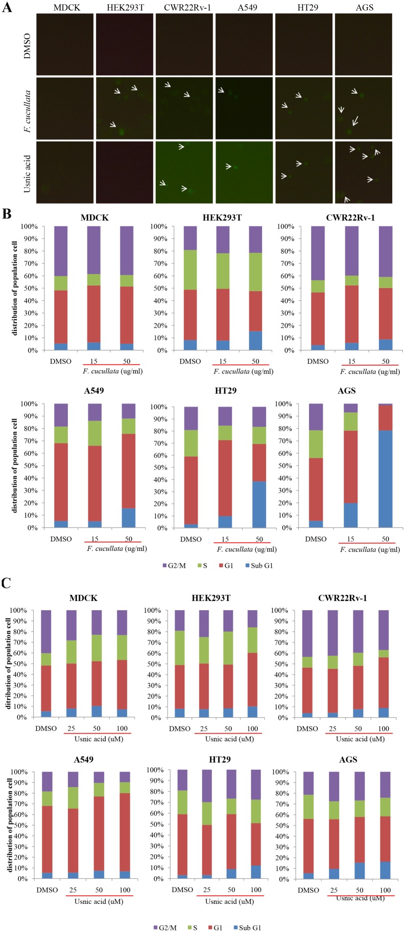
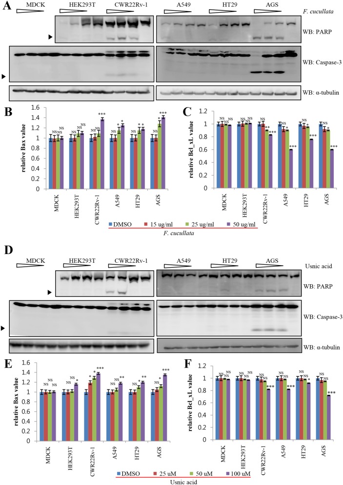
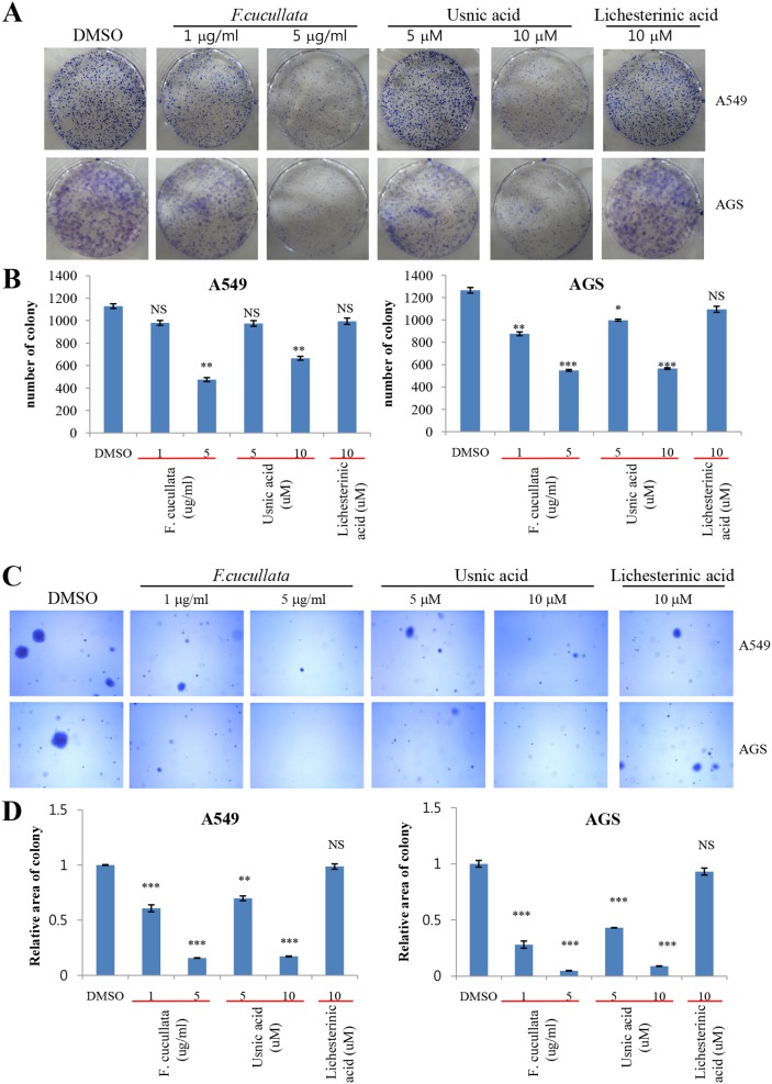
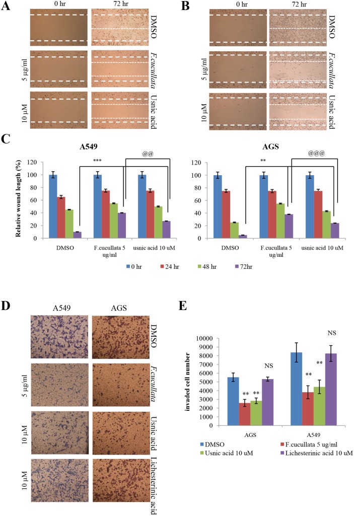
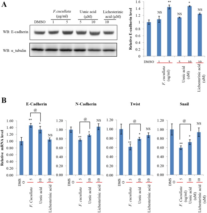
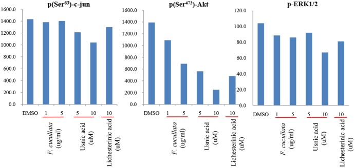
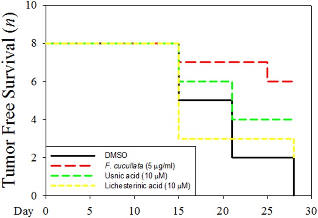
Lichens are symbiotic organisms which produce distinct secondary metabolic products. In the present study, we tested the cytotoxic activity of 17 lichen species against several human cancer cells and further investigated the molecular mechanisms underlying their anti-cancer activity. We found that among 17 lichens species, F. cucullata exhibited the most potent cytotoxicity in several human cancer cells. High performance liquid chromatography analysis revealed that the acetone extract of F. cucullata contains usnic acid, salazinic acid, Squamatic acid, Baeomycesic acid, d-protolichesterinic acid, and lichesterinic acid as subcomponents. MTT assay showed that cancer cell lines were more vulnerable to the cytotoxic effects of the extract than non-cancer cell lines. Furthermore, among the identified subcomponents, usnic acid treatment had a similar cytotoxic effect on cancer cell lines but with lower potency than the extract. At a lethal dose, treatment with the extract or with usnic acid greatly increased the apoptotic cell population and specifically activated the apoptotic signaling pathway; however, using sub-lethal doses, extract and usnic acid treatment decreased cancer cell motility and inhibited in vitro and in vivo tumorigenic potentials. In these cells, we observed significantly reduced levels of epithelial-mesenchymal transition (EMT) markers and phosphor-Akt, while phosphor-c-Jun and phosphor-ERK1/2 levels were only marginally affected. Overall, the anti-cancer activity of the extract is more potent than that of usnic acid alone. Taken together, F. cucullata and its subcomponent, usnic acid together with additional component, exert anti-cancer effects on human cancer cells through the induction of apoptosis and the inhibition of EMT.


Conflict of interest statement

Competing Interests: The authors have declared that no competing interests exist.

Figures

Figure 1
Figure 1. Cytotoxic effects of the acetone extract of F. cucullata and its main component, usnic acid, on human cancer cells.
(A) Percent viability of cells treated with the acetone extract of F. cucullata. Cells were treated with the F. cucullata extract in a concentration ranging from 10–50 µg/mL for 48 hr, and cell viability was measured by an MTT assay. (B) High performance liquid chromatography chromatograms of the F. cucullata extract. The identity of each subcomponent is noted on the corresponding peak. (C–D) The percent viability of cells treated with either usnic acid (C) or lichesterinic acid (D). Cells were treated with the indicated subcomponent of F. cucullata in a concentration ranging from 12.5–50 µM for 48 hr, and cell viability was measured by an MTT assay. Data represent means ± S.E.M. (standard error of the mean), n = 3.
Figure 2
Figure 2. Induction of nuclear condensation of human cancer cells by the acetone extract of F. cucullata and its main component, usnic acid in lethal concentrations.
(A) Hoechst 33258 staining of AGS (human gastric cancer cell line) cells treated with the F. cucullata extract or its subcomponents, usnic acid and lichesterinic acid. Arrows indicate cells showing condensed or fragmented nuclear morphology. Representative images are shown from three independent experiments. (B) Quantificational analysis of condensed or fragmented nuclear morphology in various cells treated with F. cucullata extract or its subcomponents. Data represent mean ± S.E.M. (standard error of the mean), n = 3. **p<0.01; ***p<0.001; NS, no significant difference compared to the dimethylsulfoxide-treated group.
Figure 3
Figure 3. Induction of Annexin V positivity and accumulation of sub G1 population on human cancer cells by the acetone extract of F. cucullata and usnic acid in lethal concentrations.
(A) FITC-Annexin V staining of cells treated with the F. cucullata extract or usnic acid. Arrows indicate cells showing FITC positivity. (B–C) Flow cytometric analysis of cell-cycle distributions after F. cucullata extract (B) or usnic acid (C) treatment and graphical representation of the results. Representative images or results are shown from three independent experiments.
Figure 4
Figure 4. Activation of apoptosis pathway on human cancer cells by the acetone extract of F. cucullata and usnic acid in lethal concentrations.
(A and D) Western blot analysis of poly (ADP-ribose) polymerase (PARP) and caspase-3 in cells treated with the F. cucullata (A) or usnic acid (D). Arrowheads indicate cleaved fragments of each protein. (B–C and E–F) Quantificational analysis of Bax (B and E) and Bcl-xL (C and F) protein expression levels in cells treated with the F. cucullata or usnic acid, respectively. Data represent mean ± S.E.M. (standard error of the mean). *p<0.05; **p<0.01; ***p<0.001; NS, no significant difference compared to the dimethylsulfoxide-treated group.
Figure 5
Figure 5. Inhibition of anchorage-independent growth of A549 and AGS cancer cells by the acetone extract of F. cucullata and usnic acid in sub-lethal concentrations.
(A–B) Clonogenic assay of A549 and AGS cells treated with the F. cucullata extract, usnic acid, or lichesterinic acid (A) and quantificational analysis of colony number in each group (B). (C–D) Soft agar colony-formation assay of A549 and AGS cells treated with F. cucullata extract, usnic acid, or lichesterinic acid (C) and quantificational analysis of percent colony area in each group (D). Representative images are shown from three independent experiments. Data represent mean ± S.E.M. (standard error of the mean), n = 3. *p<0.05; **p<0.01; ***p<0.001; NS, no significant difference compared to the dimethylsulfoxide-treated group.
Figure 6
Figure 6. Inhibition of the motility of A549 and AGS cancer cells by the acetone extract of F. cucullata and usnic acid in sub-lethal concentrations.
(A–C) Migration assay of A549 (A) and AGS (B) cells treated with the F. cucullata extract or usnic acid, and quantificational analysis of wound length in each group (C). (D–E) Invasion assay of A549 and AGS cells treated with the F. cucullata extract, usnic acid, or lichesterinic acid (D), and quantificational analysis of invaded cell numbers in each group (E). Representative images are shown from three independent experiments. Data represent mean ± S.E.M. (standard error of the mean), n = 3. **p<0.01; ***p<0.001; NS, no significant difference when compared to the dimethylsulfoxide-treated group in each cell lines. @@p<0.01; @@@p<0.001 when compared to the indicated group.
Figure 7
Figure 7. Suppression of epithelial-mesenchymal transition (EMT) by the acetone extract of F. cucullata and usnic acid in sub-lethal concentrations.
(A) E-cadherin level in A549 cells treated with the F. cucullata extract, usnic acid, or lichesterinic acid for 48 hr, and quantificational analysis of E-cadherin band in each group. Values were obtained by measuring the intensity of E-cadherin band normalized to α-tubulin. (B) Quantitative analysis of the mRNA level of EMT markers in A549 cells treated with the F. cucullata extract, usnic acid, or lichesterinic acid for 48 hr. Data represent mean ± S.E.M. (standard error of the mean), n = 3. *p<0.05; **p<0.01; NS, no significant difference when compared to the dimethylsulfoxide-treated group; @p<0.05 when compared to the indicated group.
Figure 8
Figure 8. Reduction of phosphor-Akt level by the acetone extract of F. cucullata and usnic acid in sub-lethal concentrations.
Phosphoprotein analysis for p(Ser63)-c-jun, p(Ser473)-Akt, and p-(Thr202/Tyr204, Thr185/Tyr187)-ERK1/2 in A549 cells treated with the F. cucullata extract, usnic acid, or lichesterinic acid.
Figure 9
Figure 9. Inhibition of in vivo tumorigenicity of A549 cancer cells pretreated by the acetone extract of F. cucullata and usnic acid in sub-lethal concentrations.
A549 cells were pretreated with indicated concentration of F. cucullata, usnic acid, or lichesterinic acid before subcutaneous injection into Balb/c nude mouse (n = 8) and tumor free survival number in each group during 4 weeks were measured.

References

    1. WHO (2013) World Health Statistics 2013.
    1. Siegel R, Naishadham D, Jemal A (2013) Cancer statistics, 2013. CA Cancer J Clin 63: 11–30. - PubMed
    1. Nash TH (2008) Lichen biology. Cambridge; New York: Cambridge University Press. ix, 486 p.
    1. Fashelt D (1994) Secondary biochemistry of lichens. Symbiosis 16: 117–165.
    1. Shrestha G, St. Clair LL (2013) Lichens: a promising source of antibiotic and anticancer drugs. Phytochem Rev 12: 229–244.

Publication types

MeSH terms

LinkOut - more resources