. 2014;5(3-4):150-9.
doi: 10.1080/17588928.2014.975680. Epub 2014 Oct 31.

Functional connectivity of the posterior hippocampus is more dominant as we age

Affiliations

Functional connectivity of the posterior hippocampus is more dominant as we age

Sonja Blum et al. Cogn Neurosci. 2014.

Abstract

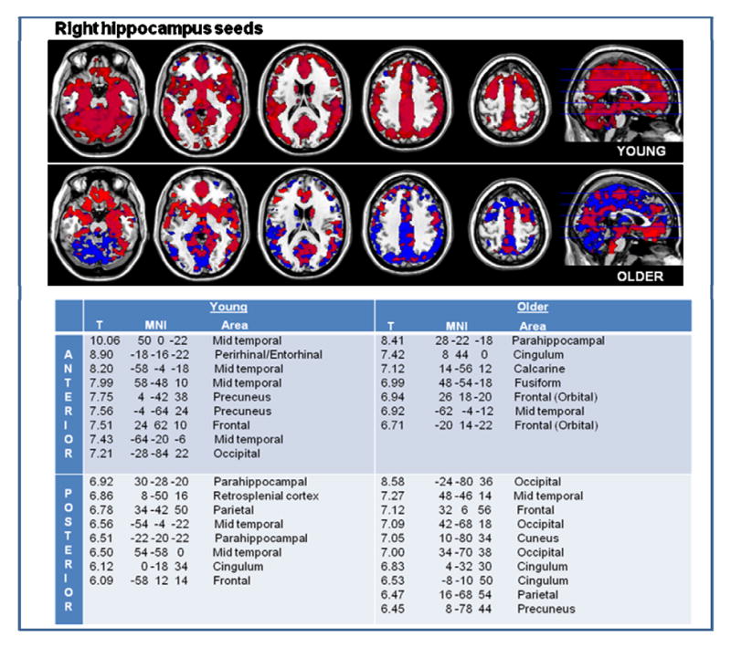
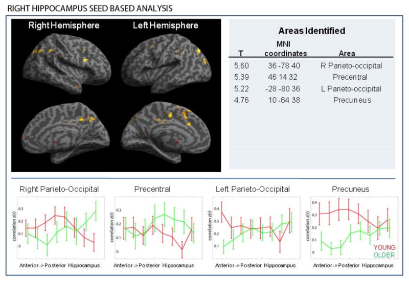
The role of the hippocampus in memory is dependent on its interaction with distributed brain areas. Anterior and posterior hippocampus have different roles in memory processing, and are impacted differently by aging in terms of structural decline, however, functional connectivity of these hippocampal regions in aging is not well understood. Young (age 17-30) and aging (age 60-69) cognitively normal subjects underwent resting-state functional MRI revealing a shift from anterior hippocampus dominant hippocampus connectivity in younger age group to posterior hippocampus dominant connectivity in aging subjects. We identified a subset of neocortical regions that are connected to the anterior hippocampus in younger adults but to the posterior hippocampus among older subjects, suggesting an age-related reorganization of hippocampal networks supporting normal cognitive function. We also performed volumetric analysis which revealed no significant structural differences between groups. These findings provide evidence that "functional anatomy" which supports normal memory performance changes across the life span.

Keywords: Hippocampus; Memory; Network.


Conflict of interest statement

There are no conflicts of interest.

Figures

Figure 1
Figure 1
Illustration of anterior (red) and posterior (blue) hippocampus segmentations, here represented on a single subject’s saggital T1 image.
Figure 2
Figure 2
In younger individuals, anterior and posterior hippocampus are widely connected to other brain areas, with areas of highest connectivity noted in the table. In the older subjects, there is overall diminished functional connectivity of the hippocampus to cortical areas, but there was a relative increase in posterior hippocampal connectivity. Anterior hippocampus functional connectivity to cortical areas is noted in red, and posterior in blue. The table lists the brain regions most connected to the anterior and those most connected to the posterior hippocampus of young and older adults.
Figure 2
Figure 2
In younger individuals, anterior and posterior hippocampus are widely connected to other brain areas, with areas of highest connectivity noted in the table. In the older subjects, there is overall diminished functional connectivity of the hippocampus to cortical areas, but there was a relative increase in posterior hippocampal connectivity. Anterior hippocampus functional connectivity to cortical areas is noted in red, and posterior in blue. The table lists the brain regions most connected to the anterior and those most connected to the posterior hippocampus of young and older adults.
Figure 3
Figure 3
A set of brain regions is preferentially connected with the anterior hippocampus in the young but to the posterior hippocampus in the older subjects. This was observed both in left and right hippocampus seed based analyses. There were no similar observations in the opposite direction (i.e. there were no areas that are preferentially connected to the posterior hippocampus in the young but to the anterior in older) (Note: results are overlaid on the inflated brain template provided in SPM 8). Largest cortical clusters identified are listed in the table. The graphs illustrate the strength of connectivity between the cortical regions and eight locations (see methods) spanning the longitudinal axis of the hippocampus. As we move from anterior to posterior hippocampus, the strength of functional connectivity with the cortical nodes decreases moving posteriorly in young subjects, whereas it increases as we move posteriorly in older subjects.
Figure 3
Figure 3
A set of brain regions is preferentially connected with the anterior hippocampus in the young but to the posterior hippocampus in the older subjects. This was observed both in left and right hippocampus seed based analyses. There were no similar observations in the opposite direction (i.e. there were no areas that are preferentially connected to the posterior hippocampus in the young but to the anterior in older) (Note: results are overlaid on the inflated brain template provided in SPM 8). Largest cortical clusters identified are listed in the table. The graphs illustrate the strength of connectivity between the cortical regions and eight locations (see methods) spanning the longitudinal axis of the hippocampus. As we move from anterior to posterior hippocampus, the strength of functional connectivity with the cortical nodes decreases moving posteriorly in young subjects, whereas it increases as we move posteriorly in older subjects.

References

    1. Arieli A, Shoham D, Hildesheim R, Grinvald A. Coherent spatiotemporal patterns of ongoing activity revealed by real-time optical imaging coupled with single-unit recording in the cat visual cortex. J Neurophysiol. 1995;73(5):2072–2093. - PubMed
    1. Behzadi Y, Restom K, Liau J, Liu TT. A component based noise correction method (CompCor) for BOLD and perfusion based fMRI. Neuroimage. 2007;37(1):90–101. doi: 10.1016/j.neuroimage.2007.04.042. - DOI - PMC - PubMed
    1. Biswal B, Yetkin FZ, Haughton VM, Hyde JS. Functional connectivity in the motor cortex of resting human brain using echo-planar MRI. Magn Reson Med. 1995;34(4):537–541. - PubMed
    1. Chen KH, Chuah LY, Sim SK, Chee MW. Hippocampal region-specific contributions to memory performance in normal elderly. Brain Cogn. 2010;72(3):400–407. doi: 10.1016/j.bandc.2009.11.007. - DOI - PubMed
    1. Dash PK, Hebert AE, Runyan JD. A unified theory for systems and cellular memory consolidation. Brain Res Brain Res Rev. 2004;45(1):30–37. doi: 10.1016/j.brainresrev.2004.02.001. - DOI - PubMed

Publication types

LinkOut - more resources