. 2014 Oct 31;3(11):1139-49.
doi: 10.1242/bio.20147500.

Dynamically-expressed prion-like proteins form a cuticle in the pharynx of Caenorhabditis elegans

Affiliations

Dynamically-expressed prion-like proteins form a cuticle in the pharynx of Caenorhabditis elegans

Julia B George-Raizen et al. Biol Open. .

Abstract

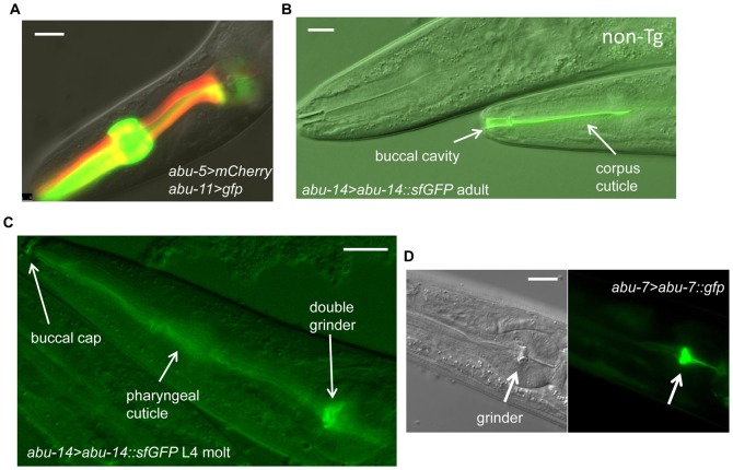
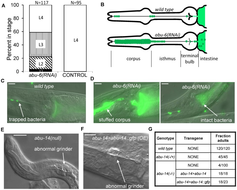
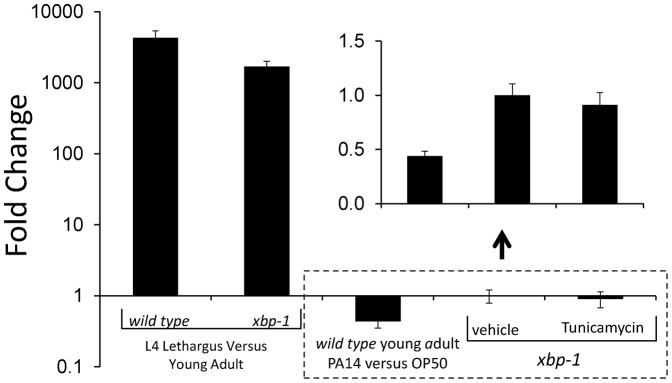
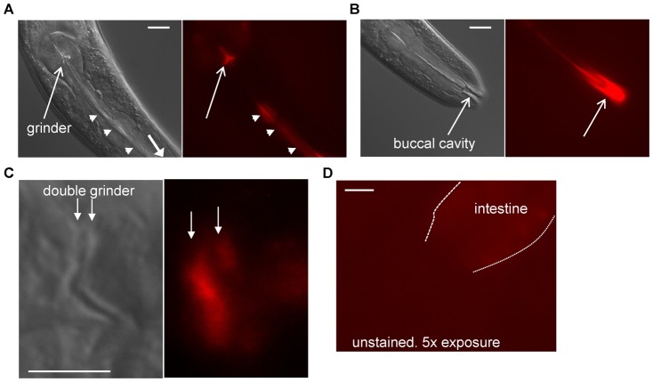
In molting animals, a cuticular extracellular matrix forms the first barrier to infection and other environmental insults. In the nematode Caenorhabditis elegans there are two types of cuticle: a well-studied collagenous cuticle lines the body, and a poorly-understood chitinous cuticle lines the pharynx. In the posterior end of the pharynx is the grinder, a tooth-like cuticular specialization that crushes food prior to transport to the intestine for digestion. We here show that the grinder increases in size only during the molt. To gain molecular insight into the structure of the grinder and pharyngeal cuticle, we performed a microarray analysis to identify mRNAs increased during the molt. We found strong transcriptional induction during the molt of 12 of 15 previously identified abu genes encoding Prion-like (P) glutamine (Q) and asparagine (N) rich PQN proteins, as well as 15 additional genes encoding closely related PQN proteins. abu/pqn genes, which we name the abu/pqn paralog group (APPG) genes, were expressed in pharyngeal cells and the proteins encoded by two APPG genes we tested localized to the pharyngeal cuticle. Deleting the APPG gene abu-14 caused abnormal pharyngeal cuticular structures and knocking down other APPG genes resulted in abnormal cuticular function. We propose that APPG proteins promote the assembly and function of a unique cuticular structure. The strong developmental regulation of the APPG genes raises the possibility that such genes would be identified in transcriptional profiling experiments in which the animals' developmental stage is not precisely staged.

Keywords: ABU/PQN; C. elegans; amyloid; cuticle; innate immunity; larvae; molting; unfolded protein response.


Conflict of interest statement

Competing interests: The authors have no competing financial interests to declare.

Figures

Fig. 1.
Fig. 1.. abu/pqn paralog group genes are up-regulated during cuticle synthesis.
(A) Pictures of a wild-type posterior pharynx at the start (0 minutes after pumping cessation) and middle (45 minutes after pumping cessation) of the fourth larval stage (L4) lethargus period. Thin and thick arrows denote the L4 and adult grinders, respectively. Anterior is to the left. Scale bar is 10 µm. (B) Width of the grinder as a function of time after hatching. First larval stage (L1) was 0–15 hours after hatching and the second larval stage was 18–26 hours after hatching. L1 lethargus occurred between 15 and 18 hours after hatching. Each point corresponds to one worm. Linear regression of the data within each larval stage produced lines with slopes that were not significantly different from zero (one-way ANOVA, p>0.1). (C) Illustration of C. elegans larval development. L1–L4 denotes larval stages 1–4, and Leth denotes lethargus. Numbers above the figure denote hours after hatching. Arrows mark sampling times for RNA collections. Dark arrows correspond to samples collected in this study whereas gray arrows correspond to sampling collected in Baugh at el. (Baugh et al., 2009). Venn diagrams illustrate logic used to define the L4 lethargus gene set and the Cuticle gene set. (D) The 13 most highly-expressed abu/pqn genes identified in all stages of cuticular synthesis. Shown is the fold change in the four comparisons. Additional genes in the abu/pqn paralog group are listed in supplementary material Table S5.
Fig. 2.
Fig. 2.. abu/pqn genes are expressed in the pharyngeal cuticle.
(A) Expression of abu-5 (red) and abu-11 (green) in pharyngeal cells. Scale bar is 10 µm. (B) Differential interference contrast (DIC) and fluorescence image of the anterior pharynx of an adult transgenic animal containing the transgene abu-14>abu-14::sfGFP. The fluorescence localizes to the pharyngeal corpus and buccal cavity cuticle (arrows). A non-transgenic sister (marked with “non-Tg”) shows no fluorescence. Anterior is to the left. Scale bar is 10 µm. (C) DIC and fluorescence image of the pharynx of a fourth larval stage molting animal containing the transgene abu-14>abu-14::sfGFP. Green fluorescence is seen in both the L4 and adult pharyngeal grinders, in the pharyngeal cuticle, and in the buccal cap. Anterior is to the left. Scale bar is 10 µm. (D) DIC (left) and fluorescence (right) images of a larval animal containing the transgene abu-7>abu-7::GFP. The pharyngeal grinder is brightly fluorescent. Scale bar is 10 µm.
Fig. 3.
Fig. 3.. Disruption of abu/pqn gene function results in cuticular abnormalities in the pharynx and poor digestion of bacteria.
(A) Developmental delay of transgenic animals expressing myo-2>abu-6(RNAi) in comparison to control transgenic animals expressing abu-5>mCherry. White, vertical, diagonal, and black bars denote, L4, L3, L2 and L1 stages, respectively. (B) Cartoon depicting observations of feeding in wild-type animals and in abu-6(RNAi) animals. Normally, bacteria (green) are trapped in the corpus and anterior isthmus, and are disrupted by the grinder in the terminal bulb to release their contents into the anterior intestine (lighter color green). In abu-6(RNAi) transgenic animals, bacteria accumulate in the corpus and are poorly disrupted by the grinder, resulting in the presence of intact bacteria in the anterior intestine. (C) Example of a wild-type animal with bacteria trapped in the anterior isthmus and with disrupted green bacteria in the anterior intestine. Anterior is to the left. Scale bar is 10 µm. (D) abu-6(RNAi) animals fed fluorescent bacteria have a pharyngeal corpus stuffed with bacteria as well as intact bacteria in the anterior intestine. Anterior is to the left. Scale bar is 10 µm. (E) Aberrant pharyngeal development observed in an abu-14(ok1789) arrested first larval stage animal. Arrow indicates abnormal grinder. Anterior is to the left. Scale bar is 10 µm. (F) Aberrant pharyngeal cuticle in an adult animal over-expressing abu-14::gfp. Arrow indicates an abnormal grinder. Anterior is to the left. Scale bar is 5 µm. (G) In the absence of abu-14, most animals do not reach adulthood 3 days after hatching. This defect is fully rescued by a genomic DNA fragment containing the abu-14 gene and is partially rescued by a DNA fragment encoding an ABU-14::GFP protein fusion expressed under the control of an abu-14 promoter.
Fig. 4.
Fig. 4.. abu/pqn genes are induced during L4 lethargus but are not induced by a blocked unfolded protein response or in response to Pseudomonas aeruginosa strain PA14 infection.
Quantitative reverse transcriptase polymerase chain reaction analysis using oligonucleotide primers that amplify abu-6, abu-7, abu-8, and abu-15. The average of four biological replicates is shown on a logarithmic scale. Error bars denote standard deviation. There is strong induction of abu-6, -7, -8 and -15 gene expression in L4 lethargus in comparison to 4-hour old adults in both wild-type and xbp-1 genetic backgrounds, but no induction in young adults in response to PA14 exposure. Treatment of xbp-1 young adults with Tunicamycin or with the vehicle 0.5% DMSO does not result in abu-6, -7, -8 and -15 gene induction. Inset shows on a magnified non-logarithmic y-axis the results of the three conditions bounded by the dotted box (the x-axis is at y = 1 on the logarithmic plot and y = 0 in the inset.).
Fig. 5.
Fig. 5.. Pharyngeal cuticle stains with amyloid dye Congo Red.
(A) DIC (left) and fluorescence (right) images that focus on the grinder (arrow) and pharyngeal lumen (arrow heads) of an animal stained with Congo Red. Anterior is down and to the right. Scale bar is 10 µm. (B) DIC (left) and fluorescence (right) images that focus on the buccal cavity (arrow) of the same animal shown in panel A. Scale bar is 10 µm. (C) DIC (left) and fluorescence (right) images that focus on the double grinder during the L4 molt. Both grinders (marked with arrows) stain with Congo Red. Anterior is to the left. Scale bar is 5 µm. (D) Camera exposure times 5 times greater than those used in panel C show no grinder fluorescence in unstained animals. Anterior is to the left and down. Scale bar is 10 µm.

References

    1. Aguinaldo A. M., Turbeville J. M., Linford L. S., Rivera M. C., Garey J. R., Raff R. A., Lake J. A. (1997). Evidence for a clade of nematodes, arthropods and other moulting animals. Nature 387, 489–493 10.1038/387489a0 - DOI - PubMed
    1. Ao W., Gaudet J., Kent W. J., Muttumu S., Mango S. E. (2004). Environmentally induced foregut remodeling by PHA-4/FoxA and DAF-12/NHR. Science 305, 1743–1746 10.1126/science.1102216 - DOI - PubMed
    1. Avery L. (1993). The genetics of feeding in Caenorhabditis elegans. Genetics 133, 897–917. - PMC - PubMed
    1. Baugh L. R., Demodena J., Sternberg P. W. (2009). RNA Pol II accumulates at promoters of growth genes during developmental arrest. Science 324, 92–94 10.1126/science.1169628 - DOI - PubMed
    1. Benjamini Y., Hochberg Y. (1995). Controlling the false discovery rate: a practical and powerful approach to multiple testing. J. R. Stat. Soc. Series B Stat. Methodol. 57, 289–300.

LinkOut - more resources