. 2015 Mar;20(3):341-9.
doi: 10.1177/1087057114557233. Epub 2014 Oct 31.

ERK and β-arrestin interaction: a converging point of signaling pathways for multiple types of cell surface receptors

Affiliations

ERK and β-arrestin interaction: a converging point of signaling pathways for multiple types of cell surface receptors

Haifeng Eishingdrelo et al. J Biomol Screen. 2015 Mar.

Abstract

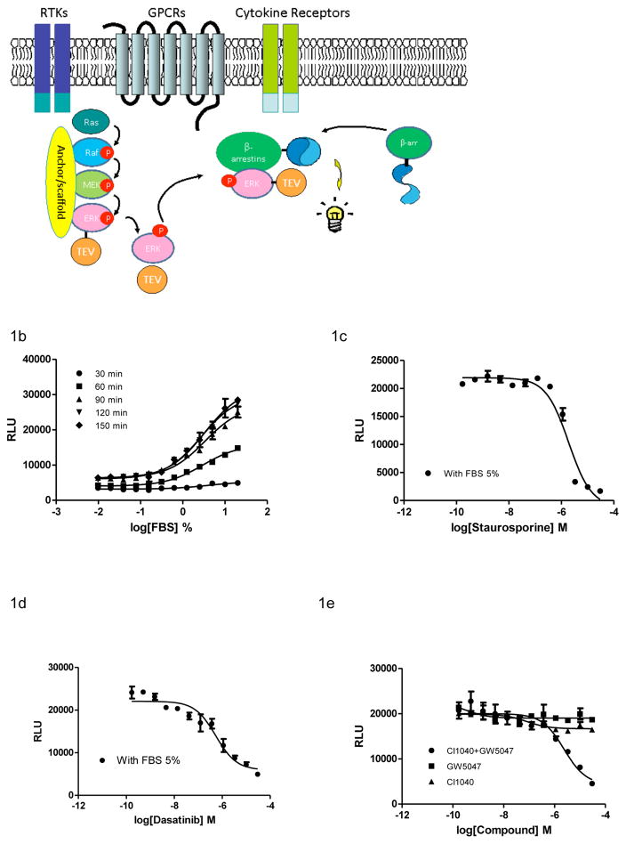
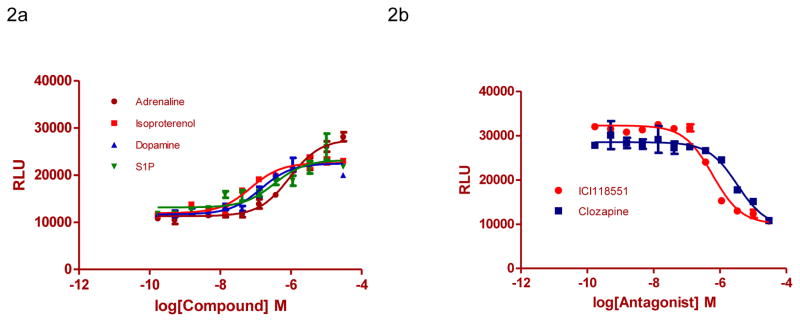
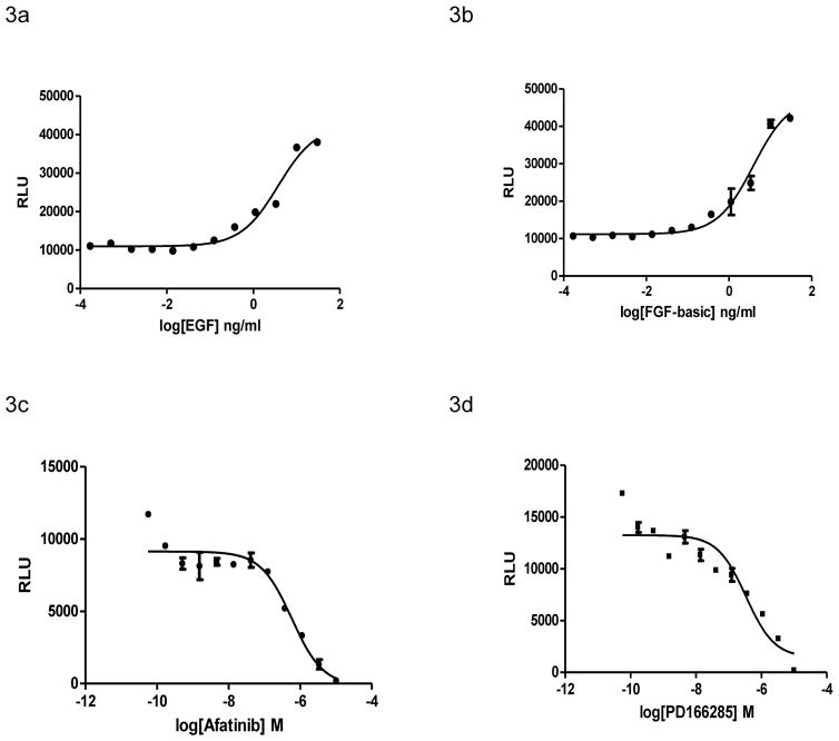
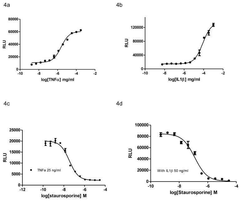
β-Arrestin, a signal adaptor protein, mediates intracellular signal transductions through protein-protein interactions by bringing two or more proteins in proximity. Extracellular signal-regulated kinase (ERK), a protein kinase in the family of mitogen-activated protein kinases (MAPKs), is involved in various receptor signal pathways. Interaction of ERK with β-arrestin or formation of ERK/β-arrestin signal complex occurs in response to activation of a variety of cell surface receptors. The ERK/β-arrestin signal complex may be a common transducer to converge a variety of extracellular stimuli to similar downstream intracellular signaling pathways. By using a cell-based protein-protein interaction LinkLight assay technology, we demonstrate a direct interaction between ERK and β-arrestin in response to extracellular stimuli, which can be sensitively and quantitatively monitored. Activations of G protein-coupled receptors (GPCRs), receptor tyrosine kinases (RTKs), and cytokine receptors promote formation of the ERK/β-arrestin signal complex. Our data indicate that the ERK/β-arrestin signal complex is a common transducer that participates in a variety of receptor signaling pathways. Furthermore, we demonstrate that receptor antagonists or kinase inhibitors can block the agonist-induced ERK and β-arrestin interaction. Thus, the ERK/β-arrestin interaction assay is useful for screening of new receptor modulators.

Keywords: ERK; extracellular signal-regulated kinases; protein-protein interactions; β-arrestin.


Conflict of interest statement

Declaration of Conflicting Interests

H.E. has the commercial interests of the patented LinkLight technology. The other authors have declared that no competing interests exist.

Figures

Figure 1
Figure 1
Design of the ERK/β-arrestin interaction LinkLight assay. (A) A schematic of a canonical RAF/MEK/ERK signaling LinkLight assay. Cell surface receptors including GPCRs, RTKs, and cytokine receptors in response to extracellular stimuli lead to activation of the RAF/MEK/ERK signaling pathway. Activated ERK realeases from the signal complex tiered by scaffold and anchor proteins and binds to β-arrestin to form ERK/β-arrestin signal complex. The close proximity between TEV-tagged ERK and pLuc-tagged β-arrestin leads to a proteolytic cleavage of permuted luciferase (pLuc). The cleaved luciferase fragments spontaneously refold to reconstitute an active luciferase, driving by the high affinity of fragment self-complementation. The luminescent signals are then detected after addition of luciferase detection reagent. (B) Dose-response curves generated using the ERK/β-arrestin interaction assay in response to different concentrations of FBS. After overnight culture, regular culture mediun was replaced with serum-free medium, and cells (20,000 cells/well in a 384-well plate) were stimulated with different concentrations of FBS for 30, 60, 90, 120, and 150 min at 37°C. (C) Dose response of an pan-kinase inhibitor staurosporine. Cells were treated with varying doses of staurospoine in serum-free DMEM for 15 min and then treated with 5% FBS for 90 min. (D) Dose response of the anti-cancer drug Dasatinib (SRC inhibitor). Cells were treated with varying doses of dasatinib in serum-free DMEM for 15 min and then treated with 5% FBS for 90 min. (E) Dose response of individual and combination of GW5074 (c-RAF inhibitor) and CI1040 (MEK inhibitor). Cells were treated with varying doses of kinase inhibitor alone or in combination in serum-free DMEM for 15 min and then stimulated with 5% FBS for 90 min. All errors bars represent SD.
Figure 2
Figure 2
ERK/β-arrestin interaction signals in response to activation of endogenous GPCRs in U2OS cells. (A) Dose-response curves generated using the ERK/β-arrestin interaction assay in response to varying doses of GPCR agonists adrenaline, isoproterenol, dopamine, and S1P. Cells were stimulated with varying doses of GPCR agonists in serum-free DMEM for 90 min at 37°C. (B) Antagonists inhibit GPCR agonist-induced ERK/β-arrestin interaction signals. Cells were treated with varying doses of GPCR antagonists BD-1047, ICI118551, and Clozapine in serum-free DMEM for 15 min and then treated with 1 μM adrenaline, isoproterenol, and dopamine respectively for 90 min.
Figure 3
Figure 3
ERK/β-arrestin interaction signals in response to activation of endogenous RTKs in U2OS cells. (A and B) Dose-response curves generated using the ERK/β-arrestin interaction LinkLight assay in response to varying doses of RTK ligands EGF and FGF-basic. Cells (20,000/well) were stimulated with varying doses of RTK ligands in serum-free DMEM for 90 min at 37°C. (C and D) Inhibition of growth factor-induced ERK/β-arrestin interaction signals. Cells were treated with varying doses of EGFR inhibitor Afatinib, and FGFR inhibitor PD166285 in serum-free DMEM for 15 min and then stimulated with 25 ng/ml EGF and FGF-basic respectively for 90 min.
Figure 4
Figure 4
ERK/β-arrestin interaction signals in response to activation of endogenous cytokine receptors in U2OS cells. (A and B) Dose-response curves generated using the ERK/β-arrestin interaction assay in response to varying doses of recombinant human TNFα and IL1β. Cells were stimulated with varying doses of TNFα and IL1β in serum-free DMEM for 90 min at 37°C. (C and D) Inhibition of TNFα and IL1β-induced signals by staurosporine. Cells were treated with varying doses of staurosporine in serum-free DMEM for 15 min and then stimulated with TNFα (25 ng/ml ) and IL1β (50 ng/ml) respectively for 90 min.

Similar articles

Cited by

References

    1. Lefkowitz RJ, Whalen EJ. beta-arrestins: traffic cops of cell signaling. Curr Opin Cell Biol. 2004;16:162–8. - PubMed
    1. Hupfeld CJ, Olefsky JM. Regulation of receptor tyrosine kinase signaling by GRKs and beta-arrestins. Annu Rev Physiol. 2007;69:561–77. - PubMed
    1. Akhtar S, Yousif MH, Chandrasekhar B, et al. Activation of EGFR/ERBB2 via pathways involving ERK1/2, P38 MAPK, AKT and FOXO enhances recovery of diabetic hearts from ischemia-reperfusion injury. PLoS One. 2012;7:e39066. - PMC - PubMed
    1. Lanner F, Rossant J. The role of FGF/Erk signaling in pluripotent cells. Development. 2010;137:3351–60. - PubMed
    1. Hwang YS, Jeong M, Park JS, et al. Interleukin-1 beta stimulates IL-8 expression through MAP kinase and ROS signaling in human gastric carcinoma cells. Oncogene. 2004;23:6603–6611. - PubMed

Publication types

MeSH terms

LinkOut - more resources