. 2014 Dec 1;42(21):13150-60.
doi: 10.1093/nar/gku1065. Epub 2014 Oct 31.

Chk2 and REGγ-dependent DBC1 regulation in DNA damage induced apoptosis

Affiliations

Chk2 and REGγ-dependent DBC1 regulation in DNA damage induced apoptosis

Martina Magni et al. Nucleic Acids Res. .

Abstract

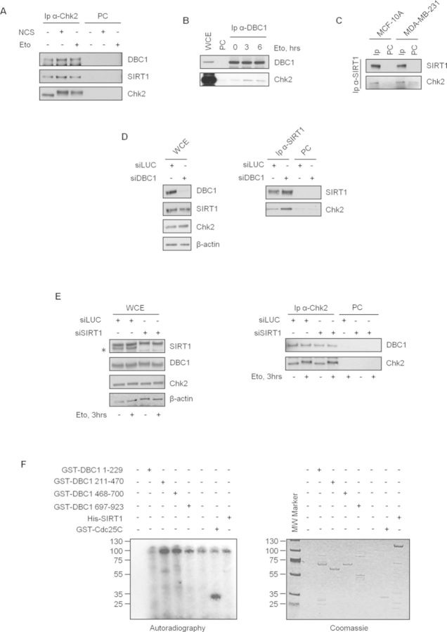
Human DBC1 (Deleted in Breast Cancer 1; KIAA1967; CCAR2) is a protein implicated in the regulation of apoptosis, transcription and histone modifications. Upon DNA damage, DBC1 is phosphorylated by ATM/ATR on Thr454 and this modification increases its inhibitory interaction with SIRT1, leading to p53 acetylation and p53-dependent apoptosis. Here, we report that the inhibition of SIRT1 by DBC1 in the DNA damage response (DDR) also depends on Chk2, the transducer kinase that is activated by ATM upon DNA lesions and contributes to the spreading of DNA damage signal. Indeed we found that inactivation of Chk2 reduces DBC1-SIRT1 binding, thus preventing p53 acetylation and DBC1-induced apoptosis. These events are mediated by Chk2 phosphorylation of the 11S proteasome activator REGγ on Ser247, which increases REGγ-DBC1 interaction and SIRT1 inhibition. Overall our results clarify the mechanisms underlying the DBC1-dependent SIRT1 inhibition and link, for the first time, Chk2 and REGγ to the ATM-DBC1-SIRT1 axis.


Figures

Figure 1.
Figure 1.
Chk2 is required for DBC1-dependent SIRT1 inhibition and induction of p53-mediated apoptosis after DNA damage in U2OS cells. Western blot analysis of total cell extracts from cells transfected with control or Chk2 siRNA and then with MOCK- or DBC1-encoding vectors. In (A) cells were incubated with 20 μM MG132 for 20 min prior to treatment with etoposide for 1 h and analyzed for p53 acetylation. Densitometric analyses (relative fold) show the ratios of acetylated p53-K382/total p53 normalized to the value of MOCK-transfected cells. In (B) and (C) cells were respectively treated with etoposide for 6 and 30 h and PUMA levels and apoptotic markers were analyzed. The same cells of experiment C were used to evaluate the percentage of dead cells by trypan blue staining. Values are mean ± SD from three independent experiments. Significant P-value is indicated (D).
Figure 2.
Figure 2.
Chk2 kinase activity promotes DBC1-SIRT1 binding in response to DNA damage in U2OS cells. DBC1 was immunoprecipitated from cells transfected with control or Chk2 siRNA and treated or not with etoposide (A) or from cells pretreated with VRX and then exposed to etoposide (B); SIRT1 presence in immunocomplexes was determined by western blot (left) and total lysates (right) were analyzed for protein levels. Relative fold indicates the densitometric quantification of SIRT1 co-immunoprecipitated with DBC1; data from etoposide-treated cells were normalized to those from untreated samples. (C) Cells were transfected with DBC1, FLAG-SIRT1 and increasing levels of HA-Chk2-encoding vectors and FLAG-SIRT1 was immunoprecipitated; immunocomplexes (left) and protein levels in total cell extracts (left) were analyzed by western blot with the indicated antibodies. (D) Cells were transfected with DBC1-WT or T454A mutant, FLAG–SIRT1- and HA-Chk2-encoding vectors and FLAG-SIRT1 was immunoprecipitated; immunocomplexes (left) and protein levels in total cell extracts (right) were analyzed by western blot with the indicated antibodies. WCE, total cell extracts; IP, immunoprecipitates; PC, pre-cleared negative control.
Figure 3.
Figure 3.
Chk2 interacts with DBC1 and SIRT1 in human cells. (AC) Co-Ip experiments demonstrating the association of Chk2 with DBC1 and SIRT1, before and after DNA damage, in different cell lines. WCE, total cell extracts; IP, immunoprecipitates; PC, pre-cleared negative control. (D) U2OS cells were transfected with control or DBC1 siRNA and endogenous SIRT1 was immunoprecipitated. Total protein levels (left) and Chk2 protein in immunocomplexes (right) were determined by western blot. (E) Chk2 was immunoprecipitated from control or siSIRT1 transfected cells before and after etoposide exposure. Total protein levels and co-Ip with DBC1 were analyzed by WB. Asterisk indicates SIRT1 specific band. (F) In vitro kinase assays with recombinant active Chk2 and GST-DBC1 deletion mutants, His SIRT1 and GST-Cdc25C (positive control) as substrates.
Figure 4.
Figure 4.
REGγ interacts with DBC1 and is phosphorylated by Chk2. (A) DBC1 is present in REGγ immunocomplexes from U2OS cells. (B) DBC1 was immunoprecipitated from U2OS cells before and after etoposide exposure. The presence of REGγ was determined by immunoblot. WCE, total cell extracts; IP, immunoprecipitates; PC, pre-cleared negative control. (C) In vitro kinase assays with recombinant active GST-Chk2 and full length or deletion mutants GST-REGγ as substrates. GST-Cdc25C was used as positive control. (D) Phos-Tag gel analyses of REGγ molecular shifts in response to etoposide and in the absence or presence of the Chk2 inhibitor VRX. (E) U2OS cells were transfected with vectors encoding FLAG-REGγWT and FLAG-REGγS247A and treated or untreated with etoposide. Molecular shifts of the ectopic proteins were analyzed by phos-tag gel.
Figure 5.
Figure 5.
REGγ mediates DBC1 and Chk2-dependent SIRT1 inhibition. (A) U2OS cells were transfected with control or REGγ siRNA, then with MOCK- or DBC1-encoding vectors and exposed to etoposide for 3 h. p53-acetylation levels were determined by western blot. (B) Control or Chk2 silenced cells overexpressing FLAG-REGγWT or FLAG-REGγS247A were expose to etoposide and analyzed for p53 acetylation. In (A) and (B) relative fold indicates the ratios of acetylated p53-K382/total p53 normalized to the value of MOCK-transfected cells. In (C) and (D) cells were transfected with MOCK-, FLAG-REGγWT- and FLAG-REGγS247A-encoding vectors treated respectively with etoposide for 6 and 30 h and PUMA levels and apoptotic markers were analyzed. (E) The same cells of experiment (D) were used to evaluate the percentage of dead cells by trypan blue staining. (F) Co-IP experiments of DBC1 and SIRT1 in REGγ-depleted cells (left) and western blot analysis of total protein levels in the same extracts (right) (G) Co-IP experiments of DBC1 and FLAG-REGγWT or FLAG-REGγS247A (left) and western blot analysis of total protein levels in the same extracts (right). Arrow indicates FLAG-positive bands.
Figure 6.
Figure 6.
Graphical representation of DBC1 regulation in the DDR. In response to DNA damage ATM contributes to p53 activation in different ways, by phosphorylation of (i) p53 on Ser15, (ii) DBC1 on Thr454 and (iii) Chk2 on Thr68. Chk2, activated by ATM, then phosphorylates REGγ, promoting DBC1-REGγ association and SIRT1 inhibition. These events finally lead to p53 activation and p53-dependent apoptosis.

Similar articles

Cited by

References

    1. Ciccia A., Elledge S.J. The DNA damage response: making it safe to play with knives. Mol. Cell. 2010;40:179–204. - PMC - PubMed
    1. Shiloh Y., Ziv Y. The ATM protein kinase: regulating the cellular response to genotoxic stress, and more. Nat. Rev. Mol. Cell Biol. 2013;14:197–210. - PubMed
    1. Stracker T.H., Usui T., Petrini J.H. Taking the time to make important decisions: the checkpoint effector kinases Chk1 and Chk2 and the DNA damage response. DNA Repair (Amst.) 2009;8:1047–1054. - PMC - PubMed
    1. Buscemi G., Zannini L., Fontanella E., Lecis D., Lisanti S., Delia D. The shelterin protein TRF2 inhibits Chk2 activity at telomeres in the absence of DNA damage. Curr. Biol. 2009;19:874–879. - PubMed
    1. Hu C., Zhang S., Gao X., Gao X., Xu X., Lv Y., Zhang Y., Zhu Z., Zhang C., Li Q., et al. Roles of kruppel-associated box (KRAB)-associated co-repressor KAP1 ser-473 phosphorylation in DNA damage response. J. Biol. Chem. 2012;287:18937–18952. - PMC - PubMed

Publication types

MeSH terms