. 2014 Nov 11;111(45):15999-6004.
doi: 10.1073/pnas.1419057111. Epub 2014 Oct 31.

Actions of the protein kinase WNK1 on endothelial cells are differentially mediated by its substrate kinases OSR1 and SPAK

Affiliations

Actions of the protein kinase WNK1 on endothelial cells are differentially mediated by its substrate kinases OSR1 and SPAK

Hashem A Dbouk et al. Proc Natl Acad Sci U S A. .

Abstract

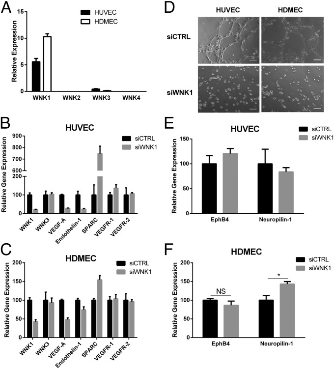
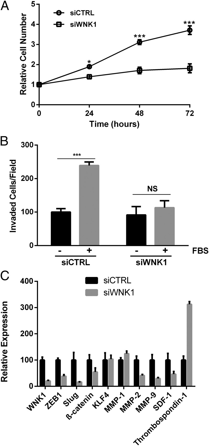
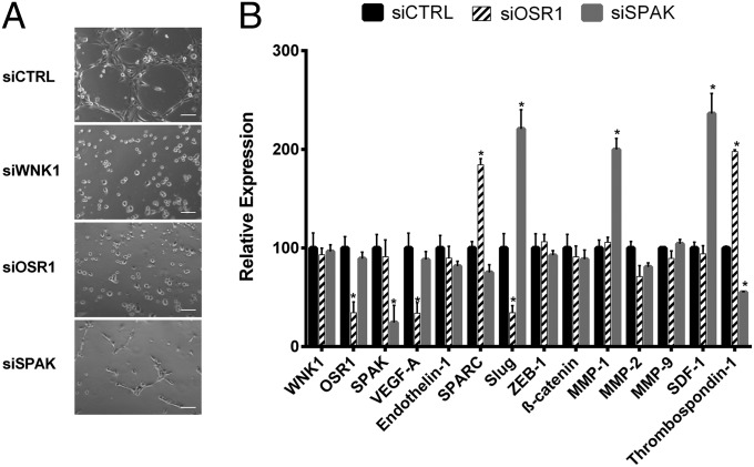
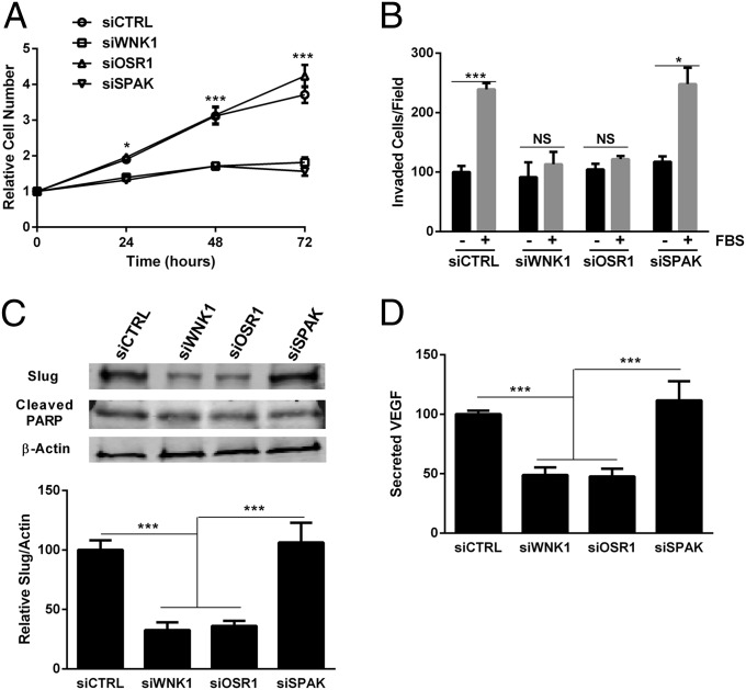
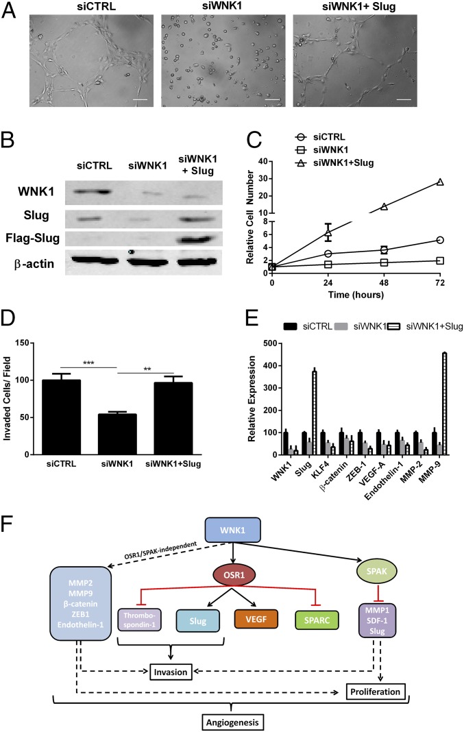
The with no lysine (K) (WNK) family of enzymes is best known for control of blood pressure through regulation of the function and membrane localization of ion cotransporters. In mice, global as well as endothelial-specific WNK1 gene disruption results in embryonic lethality due to angiogenic and cardiovascular defects. WNK1(-/-) embryos can be rescued by endothelial-specific expression of a constitutively active form of the WNK1 substrate protein kinase OSR1 (oxidative stress responsive 1). Using human umbilical vein endothelial cells (HUVECs), we explored mechanisms underlying the requirement of WNK1-OSR1 signaling for vascular development. WNK1 is required for cord formation in HUVECs, but the actions of the two major WNK1 effectors, OSR1 and its close relative SPAK (STE20/SPS1-related proline-, alanine-rich kinase), are distinct. SPAK is important for endothelial cell proliferation, whereas OSR1 is required for HUVEC chemotaxis and invasion. We also identified the zinc-finger transcription factor Slug in WNK1-mediated control of endothelial functions. Our study identifies a separation of functions for the WNK1-activated protein kinases OSR1 and SPAK in mediating proliferation, invasion, and gene expression in endothelial cells and an unanticipated link between WNK1 and Slug that is important for angiogenesis.

Keywords: EMT; Slug; angiogenesis; endothelium; migration.


Conflict of interest statement

The authors declare no conflict of interest.

Figures

Fig. 1.
Fig. 1.
WNK1 is required for cord formation in HUVECs and HDMECs. (A) Amounts of mRNAs encoding WNKs 1–4 in HUVECs and HDMECs relative to β-actin measured by qRT-PCR. (B and C) Changes in mRNAs encoding angiogenesis-related genes in (B) HUVECs and (C) HDMECs following depletion of WNK1 measured by qRT-PCR. (D) Effect of WNK1 depletion on cord formation by HUVECs and HDMECs. (Scale bars, 100 μm.) (E and F) Effect of siWNK1 on expression of venous and arterial markers in (E) HUVECs and (F) HDMECs. Data are mean ± SEM; n = 3 (AC, E, and F); representative images are from three experiments (C). *P < 0.05. NS, not significant.
Fig. 2.
Fig. 2.
WNK1 affects HUVEC proliferation and motility. (A) Proliferation of HUVECs over 72 h; cell numbers are relative to time 0. (B) Migration through a membrane toward medium without or with 10% (vol/vol) FBS. (C) Changes in mRNAs in HUVECs following depletion of WNK1 measured by qRT-PCR. Data are mean ± SEM; n = 3. *P < 0.05, ***P < 0.001.
Fig. 3.
Fig. 3.
OSR1 and SPAK impact HUVEC cord formation. (A) Effect of depletion of WNK1, OSR1, or SPAK on cord formation by HUVECs. (Scale bars, 100 μm.) (B) Changes in angiogenesis- and EMT-related genes in HUVECs following depletion of the indicated proteins. Data are mean ± SEM; n = 3 (B); representative images are from three experiments (A). *P < 0.05.
Fig. 4.
Fig. 4.
OSR1 and SPAK regulate different WNK1 functions in HUVECs. (A) Proliferation of HUVECs over 72 h. Cell numbers are relative to time 0. (B) Migration through a membrane toward medium without or with 10% FBS. (C) Lysates from HUVECs treated as indicated were immunoblotted with anti-Slug, anti-cleaved PARP, and anti–β-actin. Blots were quantified via LI-COR imaging. (D) Secreted VEGF-A was measured by ELISA following a 2-h starvation. Data are mean ± SEM; n = 3 (AC), n = 5 (D). *P < 0.05, ***P < 0.001.
Fig. 5.
Fig. 5.
Slug rescues cord formation by WNK1-depleted HUVECs. (A) Effect of Flag–Slug on cord formation by cells depleted of WNK1; representative images are from two experiments. (Scale bars, 100 μm.) (B) Lysates from HUVECs treated as indicated were immunoblotted with anti-Flag, anti-Slug, anti-WNK1, and anti–β-actin; n = 2. (C) Proliferation of cells treated as indicated over 72 h. Cell numbers are relative to time 0. (D) Migration of cells toward medium without or with 10% FBS. (E) Changes in expression of angiogenesis- and EMT-related genes in HUVECs treated as indicated. (F) Schematic diagram of the mechanisms of WNK1 actions on endothelial cell functions. Data are mean ± SD; n = 2 (CE). **P < 0.01, ***P < 0.001.

References

    1. Xu B, et al. WNK1, a novel mammalian serine/threonine protein kinase lacking the catalytic lysine in subdomain II. J Biol Chem. 2000;275(22):16795–16801. - PubMed
    1. Min X, Lee BH, Cobb MH, Goldsmith EJ. Crystal structure of the kinase domain of WNK1, a kinase that causes a hereditary form of hypertension. Structure. 2004;12(7):1303–1311. - PubMed
    1. McCormick JA, Ellison DH. The WNKs: Atypical protein kinases with pleiotropic actions. Physiol Rev. 2011;91(1):177–219. - PMC - PubMed
    1. Piala AT, et al. Chloride sensing by WNK1 involves inhibition of autophosphorylation. Sci Signal. 2014;7(324):ra41. - PMC - PubMed
    1. He G, Wang HR, Huang SK, Huang CL. Intersectin links WNK kinases to endocytosis of ROMK1. J Clin Invest. 2007;117(4):1078–1087. - PMC - PubMed

Publication types

MeSH terms