

Three-dimensional histology: tools and application to quantitative assessment of cell-type distribution in rabbit heart
- PMID: 25362175
- PMCID: PMC4217519
- DOI: 10.1093/europace/euu234
Three-dimensional histology: tools and application to quantitative assessment of cell-type distribution in rabbit heart
Abstract
Aims: Cardiac histo-anatomical organization is a major determinant of function. Changes in tissue structure are a relevant factor in normal and disease development, and form targets of therapeutic interventions. The purpose of this study was to test tools aimed to allow quantitative assessment of cell-type distribution from large histology and magnetic resonance imaging- (MRI) based datasets.
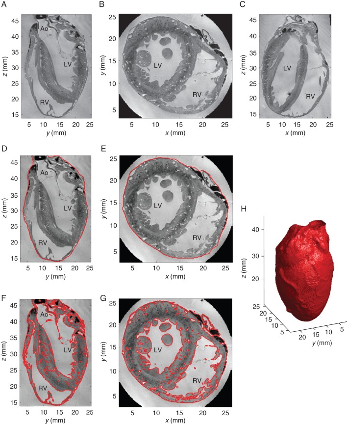
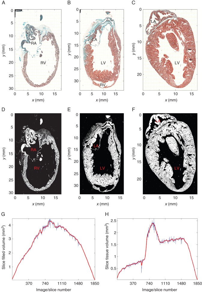
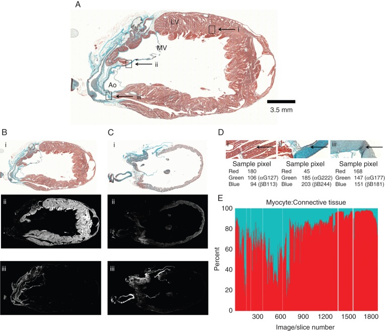
Methods and results: Rabbit heart fixation during cardioplegic arrest and MRI were followed by serial sectioning of the whole heart and light-microscopic imaging of trichrome-stained tissue. Segmentation techniques developed specifically for this project were applied to segment myocardial tissue in the MRI and histology datasets. In addition, histology slices were segmented into myocytes, connective tissue, and undefined. A bounding surface, containing the whole heart, was established for both MRI and histology. Volumes contained in the bounding surface (called 'anatomical volume'), as well as that identified as containing any of the above tissue categories (called 'morphological volume'), were calculated. The anatomical volume was 7.8 cm(3) in MRI, and this reduced to 4.9 cm(3) after histological processing, representing an 'anatomical' shrinkage by 37.2%. The morphological volume decreased by 48% between MRI and histology, highlighting the presence of additional tissue-level shrinkage (e.g. an increase in interstitial cleft space). The ratio of pixels classified as containing myocytes to pixels identified as non-myocytes was roughly 6:1 (61.6 vs. 9.8%; the remaining fraction of 28.6% was 'undefined').
Conclusion: Qualitative and quantitative differentiation between myocytes and connective tissue, using state-of-the-art high-resolution serial histology techniques, allows identification of cell-type distribution in whole-heart datasets. Comparison with MRI illustrates a pronounced reduction in anatomical and morphological volumes during histology processing.
Keywords: Cardiac MRI; Computational models; Connective tissue; Myocytes; Serial histology.
© The Author 2014. Published by Oxford University Press on behalf of the European Society of Cardiology.
Figures
Similar articles
-
Whole heart detailed and quantitative anatomy, myofibre structure and vasculature from X-ray phase-contrast synchrotron radiation-based micro computed tomography.Eur Heart J Cardiovasc Imaging. 2017 Jul 1;18(7):732-741. doi: 10.1093/ehjci/jew314. Eur Heart J Cardiovasc Imaging. 2017. PMID: 28329054
-
Magnetic resonance imaging (MRI) for the assessment of myocardial viability: an evidence-based analysis.Ont Health Technol Assess Ser. 2010;10(15):1-45. Epub 2010 Jul 1. Ont Health Technol Assess Ser. 2010. PMID: 23074392 Free PMC article.
-
2D and 3D MALDI-imaging: conceptual strategies for visualization and data mining.Biochim Biophys Acta. 2014 Jan;1844(1 Pt A):117-37. doi: 10.1016/j.bbapap.2013.01.040. Epub 2013 Mar 4. Biochim Biophys Acta. 2014. PMID: 23467008
-
Three-dimensional impulse propagation in myocardium: arrhythmogenic mechanisms at the tissue level.Circ Res. 2013 Mar 1;112(5):834-48. doi: 10.1161/CIRCRESAHA.111.300157. Circ Res. 2013. PMID: 23449546 Review.
-
Pre-clinical functional Magnetic Resonance Imaging Part II: The heart.Z Med Phys. 2014 Dec;24(4):307-22. doi: 10.1016/j.zemedi.2014.06.008. Epub 2014 Jul 9. Z Med Phys. 2014. PMID: 25023418 Review.
Cited by
-
Mapping cardiac microstructure of rabbit heart in different mechanical states by high resolution diffusion tensor imaging: A proof-of-principle study.Prog Biophys Mol Biol. 2016 Jul;121(2):85-96. doi: 10.1016/j.pbiomolbio.2016.06.001. Epub 2016 Jun 16. Prog Biophys Mol Biol. 2016. PMID: 27320383 Free PMC article.
-
Three-dimensional cardiac computational modelling: methods, features and applications.Biomed Eng Online. 2015 Apr 17;14:35. doi: 10.1186/s12938-015-0033-5. Biomed Eng Online. 2015. PMID: 25928297 Free PMC article. Review.
-
Interrogation of living myocardium in multiple static deformation states with diffusion tensor and diffusion spectrum imaging.Prog Biophys Mol Biol. 2014 Aug;115(2-3):213-25. doi: 10.1016/j.pbiomolbio.2014.08.002. Epub 2014 Aug 10. Prog Biophys Mol Biol. 2014. PMID: 25117498 Free PMC article.
-
A groupwise registration and tractography framework for cardiac myofiber architecture description by diffusion MRI: An application to the ventricular junctions.PLoS One. 2022 Jul 18;17(7):e0271279. doi: 10.1371/journal.pone.0271279. eCollection 2022. PLoS One. 2022. PMID: 35849598 Free PMC article.
-
Human-based approaches to pharmacology and cardiology: an interdisciplinary and intersectorial workshop.Europace. 2016 Sep;18(9):1287-98. doi: 10.1093/europace/euv320. Epub 2015 Nov 29. Europace. 2016. PMID: 26622055 Free PMC article.
References
-
- Katz AM, Katz PB. Homogeneity out of heterogeneity. Circulation. 1989;79:712–7. - PubMed
-
- Markhasin VS, Solovyova O, Katsnelson LB, Protsenko Y, Kohl P, Noble D. Mechano-electric interactions in heterogeneous myocardium: development of fundamental experimental and theoretical models. Prog Biophys Mol Biol. 2003;82:207–20. - PubMed
-
- Adler CP, Ringlage WP, Bohm N. [DNA content and cell number in heart and liver of children. Comparable biochemical, cytophotometric and histological investigations (author's transl)] Pathol Res Pract. 1981;172:25–41. - PubMed
-
- Camelliti P, Borg TK, Kohl P. Structural and functional characterisation of cardiac fibroblasts. Cardiovasc Res. 2005;65:40–51. - PubMed
-
- Vetter FJ, McCulloch AD. Three-dimensional analysis of regional cardiac function: a model of rabbit ventricular anatomy. Prog Biophys Mol Biol. 1998;69:157–83. - PubMed
Publication types
MeSH terms
Grants and funding
- NH/13/1/30238/BHF_/British Heart Foundation/United Kingdom
- BB/I012117/1/BB_/Biotechnology and Biological Sciences Research Council/United Kingdom
- NH/13/30238/BHF_/British Heart Foundation/United Kingdom
- PG/13/33/30210/BHF_/British Heart Foundation/United Kingdom
- FS/11/50/29038/BHF_/British Heart Foundation/United Kingdom
LinkOut - more resources
Full Text Sources
Other Literature Sources
Medical
