

A liaR deletion restores susceptibility to daptomycin and antimicrobial peptides in multidrug-resistant Enterococcus faecalis
- PMID: 25362197
- PMCID: PMC4402337
- DOI: 10.1093/infdis/jiu602
A liaR deletion restores susceptibility to daptomycin and antimicrobial peptides in multidrug-resistant Enterococcus faecalis
Abstract
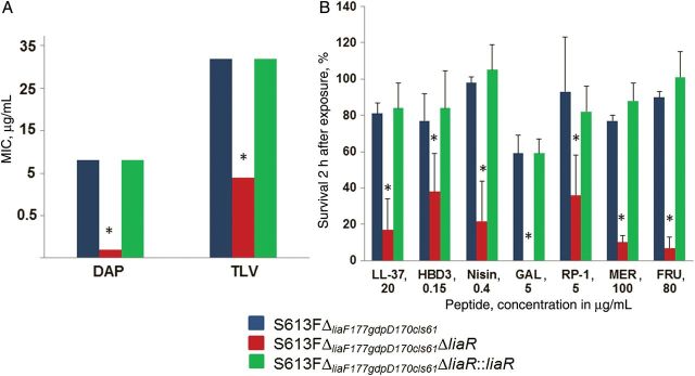
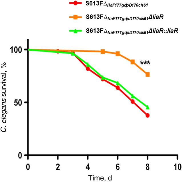
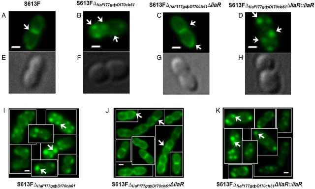
Daptomycin is a lipopeptide antibiotic that is used clinically against many gram-positive bacterial pathogens and is considered a key frontline bactericidal antibiotic to treat multidrug-resistant enterococci. Emergence of daptomycin resistance during therapy of serious enterococcal infections is a major clinical issue. In this work, we show that deletion of the gene encoding the response regulator, LiaR (a member of the LiaFSR system that controls cell envelope homeostasis), from daptomycin-resistant Enterococcus faecalis not only reversed resistance to 2 clinically available cell membrane-targeting antimicrobials (daptomycin and telavancin), but also resulted in hypersusceptibility to these antibiotics and to a variety of antimicrobial peptides of diverse origin and with different mechanisms of action. The changes in susceptibility to these antibiotics and antimicrobial peptides correlated with in vivo attenuation in a Caenorhabditis elegans model. Mechanistically, deletion of liaR altered the localization of cardiolipin microdomains in the cell membrane. Our findings suggest that LiaR is a master regulator of the enterococcal cell membrane response to diverse antimicrobial agents and peptides; as such, LiaR represents a novel target to restore the activity of clinically useful antimicrobials against these organisms and, potentially, increase susceptibility to endogenous antimicrobial peptides.
Keywords: E. faecalis; LiaFSR; antimicrobial peptides; daptomycin.
© The Author 2014. Published by Oxford University Press on behalf of the Infectious Diseases Society of America. All rights reserved. For Permissions, please e-mail: journals.permissions@oup.com.
Figures
Similar articles
-
Deletion of liaR Reverses Daptomycin Resistance in Enterococcus faecium Independent of the Genetic Background.Antimicrob Agents Chemother. 2015 Dec;59(12):7327-34. doi: 10.1128/AAC.01073-15. Epub 2015 Sep 14. Antimicrob Agents Chemother. 2015. PMID: 26369959 Free PMC article.
-
Disrupting Membrane Adaptation Restores In Vivo Efficacy of Antibiotics Against Multidrug-Resistant Enterococci and Potentiates Killing by Human Neutrophils.J Infect Dis. 2019 Jul 2;220(3):494-504. doi: 10.1093/infdis/jiz131. J Infect Dis. 2019. PMID: 30938438 Free PMC article.
-
Two Mutations Commonly Associated with Daptomycin Resistance in Enterococcus faecium LiaST120A and LiaRW73C Appear To Function Epistatically in LiaFSR Signaling.Biochemistry. 2018 Dec 11;57(49):6797-6805. doi: 10.1021/acs.biochem.8b01072. Epub 2018 Nov 27. Biochemistry. 2018. PMID: 30403130 Free PMC article.
-
Emergence and management of drug-resistant enterococcal infections.Expert Rev Anti Infect Ther. 2008 Oct;6(5):637-55. doi: 10.1586/14787210.6.5.637. Expert Rev Anti Infect Ther. 2008. PMID: 18847403 Review.
-
Multidrug-Resistant Enterococcal Infections: New Compounds, Novel Antimicrobial Therapies?Trends Microbiol. 2017 Jun;25(6):467-479. doi: 10.1016/j.tim.2017.01.004. Epub 2017 Feb 13. Trends Microbiol. 2017. PMID: 28209400 Review.
Cited by
-
Mechanism of Action and Resistance to Daptomycin in Staphylococcus aureus and Enterococci.Cold Spring Harb Perspect Med. 2016 Nov 1;6(11):a026997. doi: 10.1101/cshperspect.a026997. Cold Spring Harb Perspect Med. 2016. PMID: 27580748 Free PMC article. Review.
-
Daptomycin Resistance in Enterococcus faecium Can Be Delayed by Disruption of the LiaFSR Stress Response Pathway.Antimicrob Agents Chemother. 2021 Mar 18;65(4):e01317-20. doi: 10.1128/AAC.01317-20. Print 2021 Mar 18. Antimicrob Agents Chemother. 2021. PMID: 33468468 Free PMC article.
-
Short Antimicrobial Peptide Derived from the Venom Gland Transcriptome of Pamphobeteus verdolaga Increases Gentamicin Susceptibility of Multidrug-Resistant Klebsiella pneumoniae.Antibiotics (Basel). 2023 Dec 20;13(1):6. doi: 10.3390/antibiotics13010006. Antibiotics (Basel). 2023. PMID: 38275316 Free PMC article.
-
A variable DNA recognition site organization establishes the LiaR-mediated cell envelope stress response of enterococci to daptomycin.Nucleic Acids Res. 2015 May 19;43(9):4758-73. doi: 10.1093/nar/gkv321. Epub 2015 Apr 19. Nucleic Acids Res. 2015. PMID: 25897118 Free PMC article.
-
Mechanisms of drug resistance: daptomycin resistance.Ann N Y Acad Sci. 2015 Sep;1354:32-53. doi: 10.1111/nyas.12948. Epub 2015 Oct 23. Ann N Y Acad Sci. 2015. PMID: 26495887 Free PMC article. Review.
References
-
- World Health Organization (WHO). Antimicrobial resistance global report on surveillance 2014. Geneva, Switzerland: WHO, 2014.
-
- Centers for Diseases Control and Prevention. Antibiotic resistance threats in the United States, 2013. Atlanta, GA: Department of Health and Human Services, CDC, 2013.
-
- Muraih JK, Pearson A, Silverman J, Palmer M. Oligomerization of daptomycin on membranes. Biochim Biophys Acta 2011; 1808:1154–60. - PubMed
Publication types
MeSH terms
Substances
Grants and funding
LinkOut - more resources
Full Text Sources
Other Literature Sources
