. 2014 Dec;11(12):1253-60.
doi: 10.1038/nmeth.3170. Epub 2014 Nov 2.

A robust pipeline for rapid production of versatile nanobody repertoires

Affiliations

A robust pipeline for rapid production of versatile nanobody repertoires

Peter C Fridy et al. Nat Methods. 2014 Dec.

Abstract

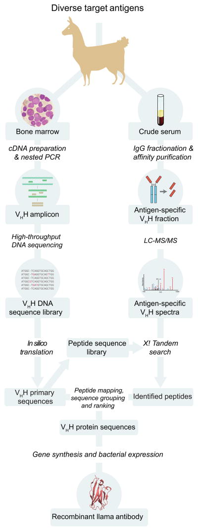
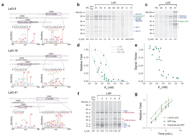
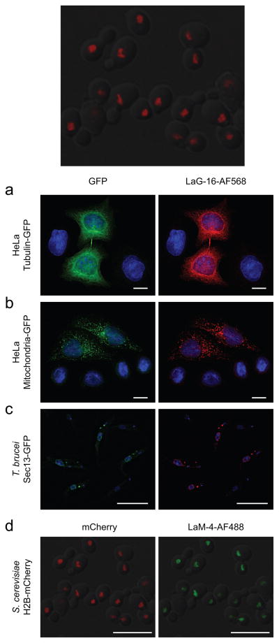
Nanobodies are single-domain antibodies derived from the variable regions of Camelidae atypical immunoglobulins. They show promise as high-affinity reagents for research, diagnostics and therapeutics owing to their high specificity, small size (∼15 kDa) and straightforward bacterial expression. However, identification of repertoires with sufficiently high affinity has proven time consuming and difficult, hampering nanobody implementation. Our approach generates large repertoires of readily expressible recombinant nanobodies with high affinities and specificities against a given antigen. We demonstrate the efficacy of this approach through the production of large repertoires of nanobodies against two antigens, GFP and mCherry, with Kd values into the subnanomolar range. After mapping diverse epitopes on GFP, we were also able to design ultrahigh-affinity dimeric nanobodies with Kd values as low as ∼30 pM. The approach presented here is well suited for the routine production of high-affinity capture reagents for various biomedical applications.


Conflict of interest statement

Competing Financial Interests:

B.T.C. and M.P.R. are inventors on a US patent application encompassing the method described in this manuscript.

Figures

Figure 1
Figure 1
Overview of nanobody identification and production pipeline. After llama immunization, cDNA from bone marrow aspirates is used for PCR amplification of the heavy-chain only variant’s variable region, which is then subjected to high-throughput DNA sequencing. Separately, the serum-derived VHH protein fraction from the same llama is affinity-purified against the antigen of interest, then analyzed by LC-MS/MS. The MS data is searched against a sequence database generated from the DNA sequencing reads, allowing identification of corresponding VHH sequences. These sequences are codon-optimized for gene synthesis, allowing efficient bacterial expression of recombinant protein. The example nanobody structure shown was obtained from PDB ID 3K1K.
Figure 2
Figure 2
MS analysis of GFP-binding VHH IgG and characterization of recombinant nanobodies. (a) Representative tandem mass spectra of identified peptides (shown boxed). Peptides were mapped to the informative CDR regions of three candidate VHH sequences, which were then chosen for production and characterization. The regions of these sequences covered by MS are underlined. (b) Indicated LaGs, commercial GFP-Trap®, or polyclonal anti-GFP llama antibody (PC) were conjugated to magnetic Dynabeads, and used for affinity isolations of S. cerevisiae Nup84-GFP or (c) RBM7-GFP from HeLa cells. Elutions were analyzed by SDS-PAGE, and duplicate Coomassie-stained bands identified by MS. Representative examples across a range of affinities are shown, and are labeled with the Kd for GFP as determined by SPR. (d) Relative yields of affinity isolated Nup84-GFP protein are plotted against the corresponding LaG’s in vitro affinity for GFP (green circles). Theoretical curves of the expected fraction of ligand bound to an immobilized binding partner at various Kds are also shown for three hypothetical ligand concentrations (grey lines). (e) The relative signal to noise ratio of three known Nup84 complex components to a known contaminant region was plotted against each LaG’s Kd. Experiments were done in duplicate, with error bars showing s.e.m. (f) S. cerevisiae mCherry-HTB2 (histone H2B) was affinity isolated by LaMs or RFP-Trap® conjugated to Dynabeads. Elutions were analyzed by SDS-PAGE, and Coomassie-stained bands identified by MS. The asterisk indicates the location of LaM nanobody leakage from the Dynabeads. LaM lanes are labeled with the Kd for mCherry as determined by SPR. (g) Affinity isolations of yeast Nup84-GFP were performed using a LaG16-LaG2 dimer with G4S, polyclonal anti-GFP, or commercial GFP- Trap®. The complex was isolated at various time points, and relative yield determined by quantification of Coomassie-stained bands of known Nup84 complex components. Experiments were done in duplicate, with error bars showing s.e.m.
Figure 3
Figure 3
Efficacy of LaG and LaM nanobodies in immunofluorescence microscopy. HeLa cells transiently transfected with (a) tubulin-emGFP or (b) an emGFP-tagged mitochondrial marker were fixed and immunostained with LaG-16 conjugated to Alexa Fluor® 568 (AF568). Cells were visualized in the green (left) and red (right) channels, with DAPI counter-staining of nuclei (blue). (c) T. brucei cells expressing GFP-tagged Sec13 were mixed 1:1 with wild-type cells, fixed, and stained with LaG-16-AF568, with DAPI counterstaining. (d) An S. cerevisiae strain with mCherry-tagged histone H2B was fixed and permeabilized, then directly stained with LaM-4 conjugated to Alexa Fluor® 488. Yeast were visualized in the red (left) and green (right) channels. All scale bars are 10 μm.
Figure 4
Figure 4
Nanobody fluorescent protein binding. (a) Thirteen high-affinity LaGs were conjugated to magnetic beads and incubated with various recombinant fluorescent proteins. All LaGs bound A. victoria (Av) GFP variants, while none bound mCherry or DsRed from Discosoma (Ds), or Phialidium (Phi) YFP. Mixed binding was observed for A. macrodactyla (Am) CFP. (b) Immobilized LaMs were similarly incubated with fluorescent proteins. All LaMs bound Discosoma mCherry, while none bound A. victoria GFP, A. macrodactyla CFP, or Phialidium YFP. Mixed binding was observed for DsRed. Example structural models were obtained from PDB IDs 1EMA (Av), 4HE4 (Phi), and 1GGX (Ds); the AmCFP model is a Phyre server prediction, .
Figure 5
Figure 5
Mapping of nanobody binding epitopes on GFP by NMR. Binding epitopes of the 11 strongest binding nanobodies on GFPuv, shown in their respective epitope group type (groups I – III). For each nanobody, two opposite sides (via a 180° rotation along a vertical axis) of the GFPuv are shown, with the binding site of the respective nanobody colored green. All GFPuv molecules are presented in space-filling mode and have the same orientation in all panels. The 3 panels on the lowest right show the GFP-Trap® nanobody (top) binding epitope and dimerization site (middle) on GFPuv as well as its ribbon diagram depicting the secondary structure elements (bottom).

References

    1. Cristea IM, Williams R, Chait BT, Rout MP. Fluorescent proteins as proteomic probes. Molecular & cellular proteomics: MCP. 2005;4:1933–1941. - PubMed
    1. Rigaut G, et al. A generic protein purification method for protein complex characterization and proteome exploration. Nature biotechnology. 1999;17:1030–1032. - PubMed
    1. Ho Y, et al. Systematic identification of protein complexes in Saccharomyces cerevisiae by mass spectrometry. Nature. 2002;415:180–183. - PubMed
    1. Domanski M, et al. Improved methodology for the affinity isolation of human protein complexes expressed at near endogenous levels. Biotechniques. 2012;0:1–6. - PMC - PubMed
    1. Gingras AC, Aebersold R, Raught B. Advances in protein complex analysis using mass spectrometry. J Physiol. 2005;563:11–21. - PMC - PubMed

Publication types

MeSH terms