. 2014 Dec;17(12):1673-81.
doi: 10.1038/nn.3858. Epub 2014 Nov 2.

Glial ankyrins facilitate paranodal axoglial junction assembly

Affiliations

Glial ankyrins facilitate paranodal axoglial junction assembly

Kae-Jiun Chang et al. Nat Neurosci. 2014 Dec.

Abstract

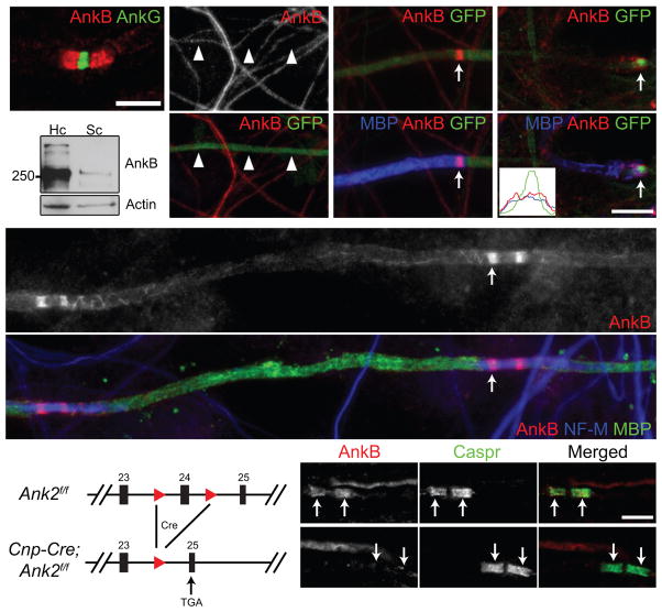
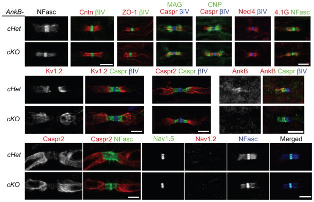
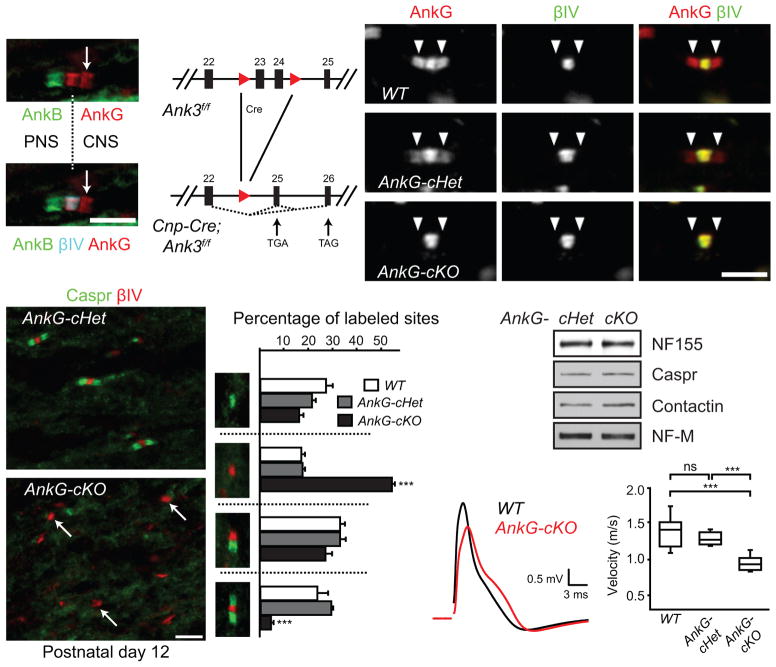
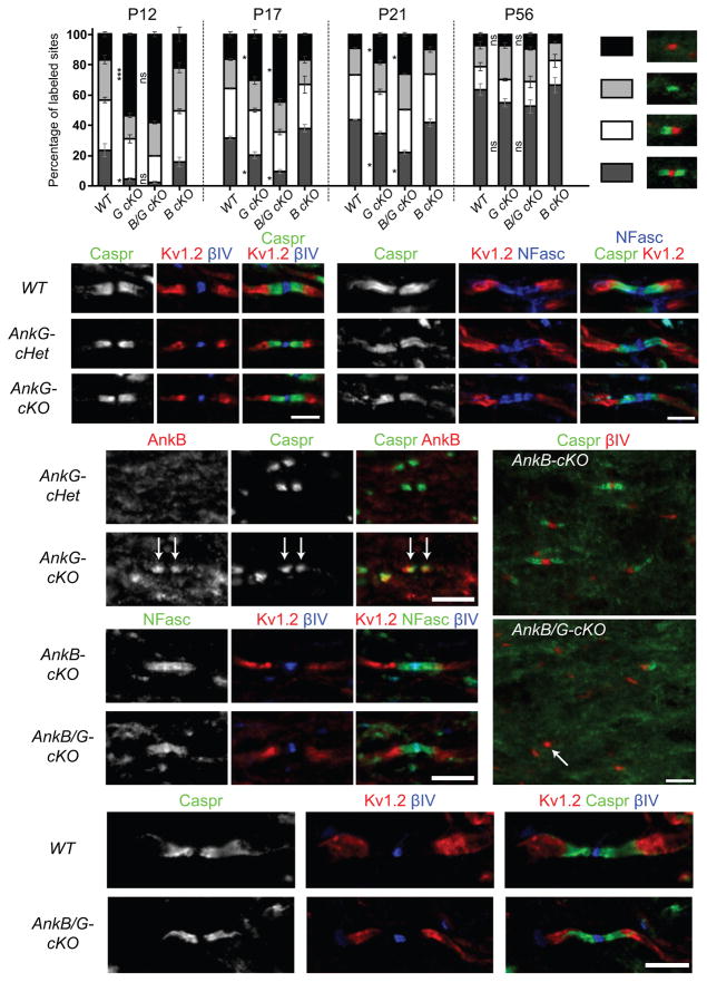
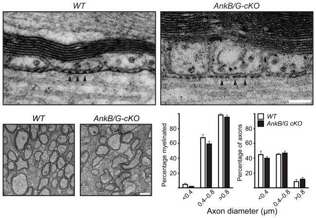
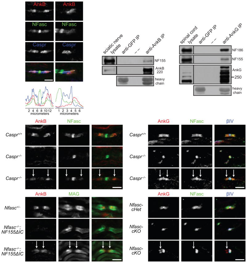
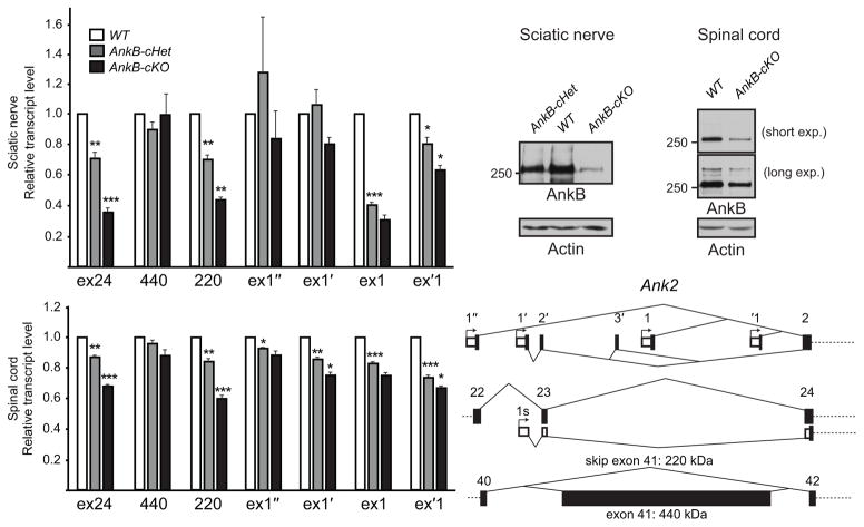
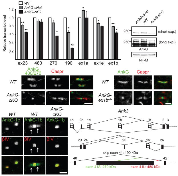
Neuron-glia interactions establish functional membrane domains along myelinated axons. These include nodes of Ranvier, paranodal axoglial junctions and juxtaparanodes. Paranodal junctions are the largest vertebrate junctional adhesion complex, and they are essential for rapid saltatory conduction and contribute to assembly and maintenance of nodes. However, the molecular mechanisms underlying paranodal junction assembly are poorly understood. Ankyrins are cytoskeletal scaffolds traditionally associated with Na(+) channel clustering in neurons and are important for membrane domain establishment and maintenance in many cell types. Here we show that ankyrin-B, expressed by Schwann cells, and ankyrin-G, expressed by oligodendrocytes, are highly enriched at the glial side of paranodal junctions where they interact with the essential glial junctional component neurofascin 155. Conditional knockout of ankyrins in oligodendrocytes disrupts paranodal junction assembly and delays nerve conduction during early development in mice. Thus, glial ankyrins function as major scaffolds that facilitate early and efficient paranodal junction assembly in the developing CNS.


Conflict of interest statement

Competing Financial Interests:

The authors declare no competing financial interests.

Figures

Figure 1
Figure 1
Paranodal AnkB is derived from Schwann cells in the PNS. (a) Immunostaining of a mouse sciatic nerve for AnkG (node, rabbit polyclonal anti-AnkG) and AnkB (paranodes, N105/17). (b) Cultured DRG neurons were infected with adenovirus carrying a GFP and AnkB shRNA-expressing construct, and immunostained (AnkB, N105/13). Arrowheads point to the GFP-positive axon. (c, d) Schwann cells were added to the same culture as in (b) and induced to myelinate. The co-culture was labeled for myelin basic protein (MBP), GFP and AnkB (N105/13 (c) or N105/17 (d)). The arrows point to paranodal AnkB. A line scan of fluorescence intensity of the paranode indicated in (d) is shown in the inset. (e) Immunoblots of lysates from rat hippocampal (Hc) neuron and purified Schwann cell (Sc) cultures (AnkB, N105/17). The full blots are presented in Supplementary Fig. 3. (f) DRG neurons from the AnkB conventional KO were co-cultured with myelinating rat Schwann cells and immunostained for AnkB (N105/17), neurofilament-M (NF-M) and MBP. The arrow points to a paranode. Localization of AnkB along the inner mesaxon was also observed as spiral extensions from paranodal junctions. (g) The scheme of the Ank2 conditional allele. The two loxP sites (red triangles) flank exon 24. After Cre recombination and removal of exon 24, a premature stop codon is generated in exon 25. (h, i) Immunostaining of 4-week-old AnkB-cHet (h) and AnkB-cKO (i) sciatic nerves (AnkB, rabbit polyclonal anti-AnkB). Arrows point to paranodal junctions. Scale bars = 5 μm (a; h, i), and 10 μm (b–d, f).
Figure 2
Figure 2
No paranodal or axonal abnormalities were observed in sciatic nerves of AnkB-cKO mice. (a–l) Immunostaining of sciatic nerve longitudinal sections from 4 to 6-week-old (a–i, l), P3 (j, AnkB, N105/17) and 1-year-old (k) mice shows normal paranodal and juxtaparanodal domains and proper Nav channel subtype switch in AnkB-cKO mice. NFasc, neurofascin (both nodal 186-kDa and paranodal 155-kDa isoforms were stained). Cntn, contactin. βIV, βIV spectrin. ZO-1, zona occludens protein 1. MAG, myelin-associated glycoprotein. CNP, 2′,3′-cyclic nucleotide 3′ phosphodiesterase. Necl4, nectin-like protein 4. Two mice per genotype and more than 75 (j) or 150 (a–i, k, l) nodes were examined in each mouse. Scale bars = 5 μm (a, b; c–g; h; i; j; k; l).
Figure 3
Figure 3
Paranodal junction assembly is disrupted in AnkG-cKO mice. (a) The dorsal root entry zone from a P7 mouse spinal cord immunostained for AnkB (N105/13), AnkG (N106/36) and βIV spectrin. The dotted line indicates the transition from PNS to CNS. The arrow points to the CNS paranode. (b) The scheme of the Ank3 conditional allele. The two loxP sites (red triangles) flank exons 23 and 24. After Cre recombination, a premature stop codon is generated in exon 25 or 26. (c–e) Immunostaining of P22 optic nerves (AnkG, N106/65). Arrowheads point to paranodes. (f, g) Immunostaining of P12 optic nerves. Arrows point to three nodal clusters without accompanying paranodal junctions. (h) Percentages of full nodes and nodal intermediates in P12 optic nerves (N = 3 for all genotypes). Unpaired two-tailed t tests (cKO vs. cHet): nodes alone: p = 0.000003; full nodes: p = 0.00001. The bar graphs show mean + SEM. ***, p < 0.001. (i) Immunoblots of P12 optic nerve homogenates. The full blots are presented in Supplementary Fig. 4. (j, k) Conduction of P17 optic nerves was measured using suction electrodes. N = 8 for WT and AnkG-cKO; N = 6 for AnkG-cHet. Two-tailed Mann-Whitney tests: WT vs. cHet: p = 0.282; WT vs. cKO: p = 0.0003; cHet vs. cKO: p = 0.0007. Data are shown in box-and-whisker plots (maximum, 75th percentile, median, 25th percentile, and minimum are shown). ***, p < 0.001; ns, not significant. Scale bars = 5 μm (a; f, g); 3 μm (c–e).
Figure 4
Figure 4
Paranodal junctions recover over time in the CNS of the AnkG-cKO and AnkB/G-cKO mice. (a) Percentages of full nodes and nodal intermediates in P12, P17, P21 and P56 optic nerves. WT, wild type. G cKO, AnkG-cKO. B cKO, AnkB-cKO. B/G cKO, AnkB/G-cKO. The data for P12 WT and G cKO are the same as those shown in Fig. 3h. N = 2 mice for AnkB/G-cKO at P12, AnkB-cKO at P17 and all the genotypes at P21; N = 3 mice for the others. 200–500 sites per animal at P12 and 600–1400 at P17–P56 were counted. Unpaired two-tailed t tests: ***, p < 0.001; *, p < 0.05; ns, not significant (for exact p values, please see the supplementary methods checklist). The bar graphs show mean ± SEM. (b–k) Immunostaining of optic nerves from P22 (b, d, e), 1-year-old (c, j, k), P12 (f, i) and P21 (g, h) mice with antibodies as indicated. The anti-AnkB N105/17 was used in (d, e). The arrows in (e) point to paranodal AnkB; the arrow in (i) points to one nodal cluster of βIV spectrin without accompanying paranodal junctions. At least two mice per genotype and more than 150 nodes of each mouse were examined. Scale bars = 3 μm (b; c), and 5 μm (d–e; f, i; g–h; j–k).
Figure 5
Figure 5
Paranodal junction assembly and myelination proceed in the absence of both AnkB and AnkG in oligodendrocytes. (a) Electron micrographs of adult optic nerve longitudinal sections. Arrowheads point to the transverse bands. (b) Electron micrographs of P17 optic nerve cross sections. (c) Percentage of axons in each diameter range that were myelinated. (d) Percentage of total axons that belonged to different groups of diameter ranges. N = 3 for each genotype shown in (c, d). The bar graphs show mean + SEM. Scale bars = 100 nm (a) and 1 μm (b).
Figure 6
Figure 6
AnkB and AnkG interact with NF155 in vivo and can be targeted to paranodes independently of paranodal junctions and NF155. (a–d) P8 sciatic nerve sections were stained for AnkB (N105/17), NFasc and Caspr (a, b), and the respective longitudinal line scans are shown (c, d). (e) Immunoprecipitation of AnkB from adult rat sciatic nerves co-precipitated NF155. Immunoprecipitation with the anti-GFP antibody served as a negative control. Heavy chain, mouse IgG heavy chain. AnkB was detected by H-300 rabbit polyclonal antibodies. (f) Immunoprecipitation of AnkG from P21 mouse spinal cords co-precipitated NF186 and NF155. AnkG was detected by the goat polyclonal antibodies. The immunoprecipitation in (e, f) was reproduced at least three times. The full blots are presented in Supplementary Fig. 3. (g–i) Immunostaining of P7 sciatic nerves (AnkB, rabbit polyclonal). Arrows point to the residual AnkB and NFasc at paranodes. (j–l) Immunostaining of P7 spinal cords (AnkG, N106/36). Arrows point to the residual AnkG and NFasc at paranodes. (m–o) Immunostaining of P5 sciatic nerves (AnkB, rabbit polyclonal). Arrows point to the paranodes with residual AnkB. (p–r) Immunostaining of P12 Nfasc-cHet (Cnp-Cre;Nfascf/+) and Nfasc-cKO (Cnp-Cre;Nfascf/f) spinal cords (AnkG, N106/36). Two mice per genotype and more than 100 nodes were examined in each mouse. 80–190 nodes per animal were quantified. Scale bars = (a, b) 5 μm for (a) and 3.3 μm for (b); 5 μm (g–i, j–l, m–o, and p–r).
Figure 7
Figure 7
Schwann cells and oligodendrocytes express 220-kDa isoform of AnkB from multiple promoters. (a) RT-qPCR analysis of P31 sciatic nerves shows the transcript levels relative to WT after normalization to Polr2a as the internal control. N = 4 for all genotypes analyzed. Unpaired two-tailed t tests (1 vs. cHet, one-sample; cHet vs. cKO, two-sample, respectively): *, p < 0.05; **, p < 0.01; ***, p < 0.001 (for exact p values, please see the supplementary methods checklist). The bar graphs show mean + SEM. (b) Immunoblots of adult sciatic nerve homogenates probed for AnkB (H-300 rabbit antibody) and actin. (c) Immunoblots of membrane homogenates from P42 spinal cords probed for AnkB (N105/17) and actin. The full blots in (b, c) are presented in Supplementary Fig. 4. (d) RT-qPCR analysis of P31 spinal cords shows the transcript levels relative to WT after normalization to Polr2a as the internal control. N = 4 for all genotypes analyzed. Unpaired two-tailed t tests (1 vs. cHet, one-sample; cHet vs. cKO, two-sample, respectively): *, p < 0.05; **, p < 0.01; ***, p < 0.001 (for exact p values, please see the supplementary methods checklist). The bar graphs show mean + SEM. (e) The schema shows the alternative first exons from different promoters and alternative splicing of exon 41 of Ank2.
Figure 8
Figure 8
Oligodendrocytes express the 270-kDa and 190-kDa isoforms of AnkG from the promoter upstream to exon 1b. (a) RT-qPCR analysis of P31 spinal cords shows the transcript levels relative to WT after normalization to Polr2a as the internal control. N = 3 for all genotypes analyzed. Unpaired two-tailed t tests (1 vs. cHet, one-sample; cHet vs. cKO, two-sample, respectively): *, p < 0.05; **, p < 0.01 (for exact p values, please see the supplementary methods checklist). The bar graphs show mean + SEM. (b) Immunoblots of membrane homogenates from P31 spinal cords probed for AnkG (goat polyclonal antibody) and NF-M. The full blots are presented in Supplementary Fig. 4. (c) Immunostaining of P12 spinal cords with antibodies against 480/270-kDa AnkG isoforms and Caspr. (d) Immunostaining of P22 spinal cords with antibodies against the peptide sequence encoded by Ank3 exon 1e or 1b and βIV spectrin. Arrows point to paranodes. (e) P90 brain stems were labeled for AnkG (N106/36) and Caspr. (f) The schema shows the alternative first exons from different promoters and alternative splicing of exon 41 of Ank3. Scale bars = 5 μm (c), 3 μm (d), and 2.5 μm (e).

References

    1. Poliak S, Peles E. The local differentiation of myelinated axons at nodes of Ranvier. Nat Rev Neurosci. 2003;4:968–980. - PubMed
    1. Salzer JL. Polarized domains of myelinated axons. Neuron. 2003;40:297–318. - PubMed
    1. Babbs CF, Shi R. Subtle paranodal injury slows impulse conduction in a mathematical model of myelinated axons. PLoS One. 2013;8:e67767. - PMC - PubMed
    1. Chang KJ, Rasband MN. Excitable domains of myelinated nerves: axon initial segments and nodes of Ranvier. Curr Top Membr. 2013;72:159–192. - PubMed
    1. Bennett V, Lorenzo DN. Spectrin- and ankyrin-based membrane domains and the evolution of vertebrates. Curr Top Membr. 2013;72:1–37. - PubMed

Publication types

LinkOut - more resources