

Endothelial responses of magnesium and other alloying elements in magnesium-based stent materials
- PMID: 25363018
- PMCID: PMC4873155
- DOI: 10.1039/c4mt00244j
Endothelial responses of magnesium and other alloying elements in magnesium-based stent materials
Abstract
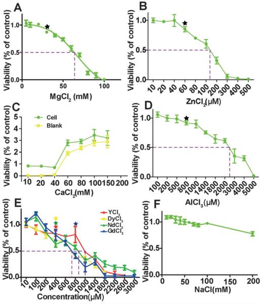
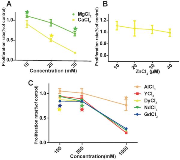
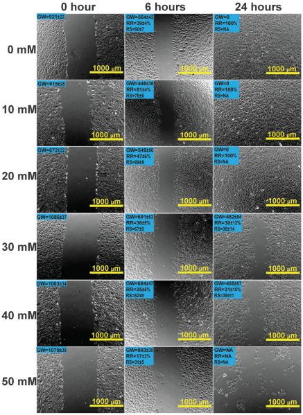
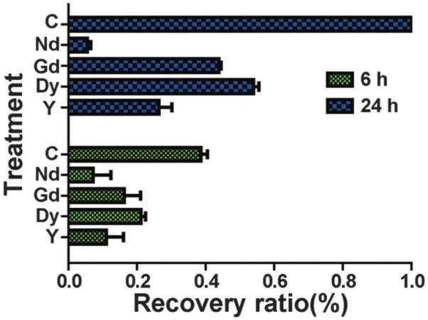
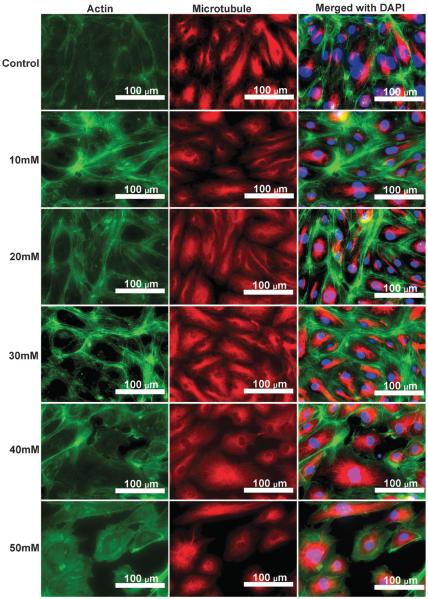
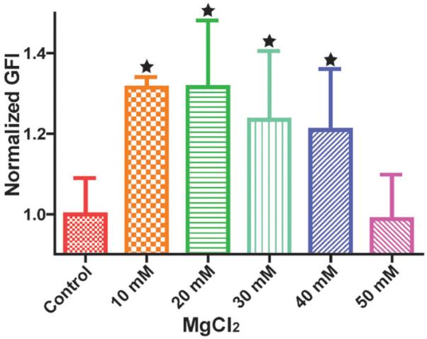
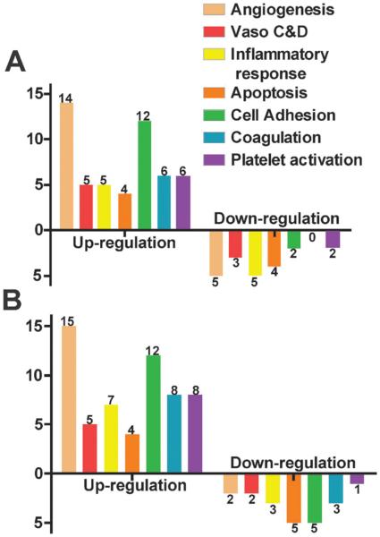
Biodegradable tailored magnesium (Mg) alloys are some of the most promising scaffolds for cardiovascular stents. During the course of degradation after implantation, all the alloying elements in the scaffold will be released to the surrounding vascular tissues. However, fundamental questions regarding the toxicity of alloying elements towards vascular cells, the maximum amount of each element that could be used in alloy design, or how each of the alloying elements affects vascular cellular activity and gene expression, are still not fully answered. This work systematically addressed these questions by revealing how application of different alloying elements commonly used in Mg stent materials influences several indices of human endothelial cell health, i.e., viability, proliferations, cytoskeletal reorganizations, migration, and the gene expression profile. The overall cell viability and proliferation showed a decreasing trend with increasing concentrations of the ions, and the half maximal effective concentrations (EC50) for each element were determined. When applied at a low concentration of around 10 mM, Mg had no adverse effects but improved cell proliferation and migration instead. Mg ions also altered endothelial gene expression significantly in a dose dependent manner. Most of the changed genes are related to angiogenesis and the cell adhesion signaling pathways. Findings from this work provide useful information on maximum safe doses of these ions for endothelial cells, endothelial responses towards these metal ions, and some guidance for future Mg stent design.
Figures

Similar articles
-
Nanophasic biodegradation enhances the durability and biocompatibility of magnesium alloys for the next-generation vascular stents.Nanoscale. 2013 Oct 21;5(20):9517-22. doi: 10.1039/c3nr02912c. Nanoscale. 2013. PMID: 23989064
-
Comparative in vitro study on binary Mg-RE (Sc, Y, La, Ce, Pr, Nd, Sm, Eu, Gd, Tb, Dy, Ho, Er, Tm, Yb and Lu) alloy systems.Acta Biomater. 2020 Jan 15;102:508-528. doi: 10.1016/j.actbio.2019.11.013. Epub 2019 Nov 10. Acta Biomater. 2020. PMID: 31722254
-
In vitro biocompatibility and endothelialization of novel magnesium-rare Earth alloys for improved stent applications.PLoS One. 2014 Jun 12;9(6):e98674. doi: 10.1371/journal.pone.0098674. eCollection 2014. PLoS One. 2014. PMID: 24921251 Free PMC article.
-
Degradable magnesium-based alloys for biomedical applications: The role of critical alloying elements.J Biomater Appl. 2019 May;33(10):1348-1372. doi: 10.1177/0885328219834656. Epub 2019 Mar 9. J Biomater Appl. 2019. PMID: 30854910 Review.
-
[Corrosive degradation of magnesium and its alloy as endovascular stent].Sheng Wu Yi Xue Gong Cheng Xue Za Zhi. 2011 Dec;28(6):1246-50. Sheng Wu Yi Xue Gong Cheng Xue Za Zhi. 2011. PMID: 22295723 Review. Chinese.
Cited by
-
Magnesium Regulates Endothelial Barrier Functions through TRPM7, MagT1, and S1P1.Adv Sci (Weinh). 2019 Jul 30;6(18):1901166. doi: 10.1002/advs.201901166. eCollection 2019 Sep 18. Adv Sci (Weinh). 2019. PMID: 31559137 Free PMC article.
-
The Flow-Induced Degradation and Vascular Cellular Response Study of Magnesium-Based Materials.Front Bioeng Biotechnol. 2022 Jul 7;10:940172. doi: 10.3389/fbioe.2022.940172. eCollection 2022. Front Bioeng Biotechnol. 2022. PMID: 35875490 Free PMC article.
-
Benchtop proof of concept and comparison of iron- and magnesium-based bioresorbable flow diverters.J Neurosurg. 2022 Dec 16;139(1):150-156. doi: 10.3171/2022.11.JNS222213. Print 2023 Jul 1. J Neurosurg. 2022. PMID: 36681964 Free PMC article.
-
The biological responses and mechanisms of endothelial cells to magnesium alloy.Regen Biomater. 2021 May 28;8(3):rbab017. doi: 10.1093/rb/rbab017. eCollection 2021 Jun. Regen Biomater. 2021. PMID: 34211729 Free PMC article.
-
Cytocompatibility and early inflammatory response of human endothelial cells in direct culture with Mg-Zn-Sr alloys.Acta Biomater. 2017 Jan 15;48:499-520. doi: 10.1016/j.actbio.2016.10.020. Epub 2016 Oct 13. Acta Biomater. 2017. PMID: 27746360 Free PMC article.
References
-
- Mao L, Yuan GY, Niu JL, Zong Y, Ding WJ. Mater. Sci. Eng., C. 2013;33:242–250. - PubMed
-
- Ge Q, Dellasega D, Demir AG, Vedani M. Acta Biomater. 2013;9.10:8604–8610. - PubMed
-
- Fan J, Qiu X, Niu X, Tian Z, Sun W, Liu X, Li Y, Li W, Meng J. Mater. Sci. Eng., C. 2013;33.4:2345–2352. - PubMed
-
- Bornapour M, Muja N, Shum-Tim D, Cerruti M, Pekguleryuz M. Acta Biomater. 2013;9:5319–5330. - PubMed
-
- Li Z, Gu X, Lou S, Zheng Y. Biomaterials. 2008;29:1329–1344. - PubMed
Publication types
MeSH terms
Substances
Grants and funding
LinkOut - more resources
Full Text Sources
Other Literature Sources
