

Ultrasound imaging in the diagnosis of benign and suspicious adrenal lesions
- PMID: 25363391
- PMCID: PMC4228948
- DOI: 10.12659/MSM.890800
Ultrasound imaging in the diagnosis of benign and suspicious adrenal lesions
Abstract
Background: The purpose of this study was to define the ultrasound imaging characteristics of adrenal tumors and to assess the performance of ultrasound in differentiating benign 'leave-alone' lesions from suspicious lesions.
Material and methods: We enrolled 882 patients in this study. The nature of each lesion was determined by histopathology. Ultrasound finding of each lesion was compared with its corresponding histopathologic result. The final study group consisted of 911 adrenal masses in 882 patients. All images were reviewed by 2 experienced investigators in a double blind manner.
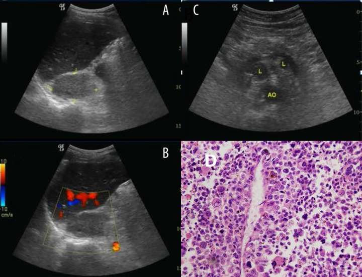
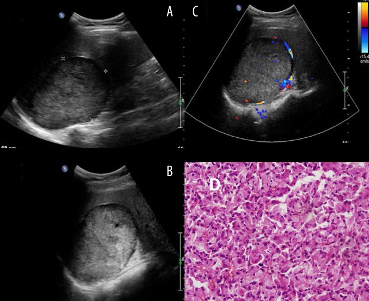
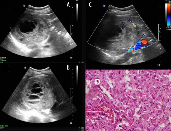
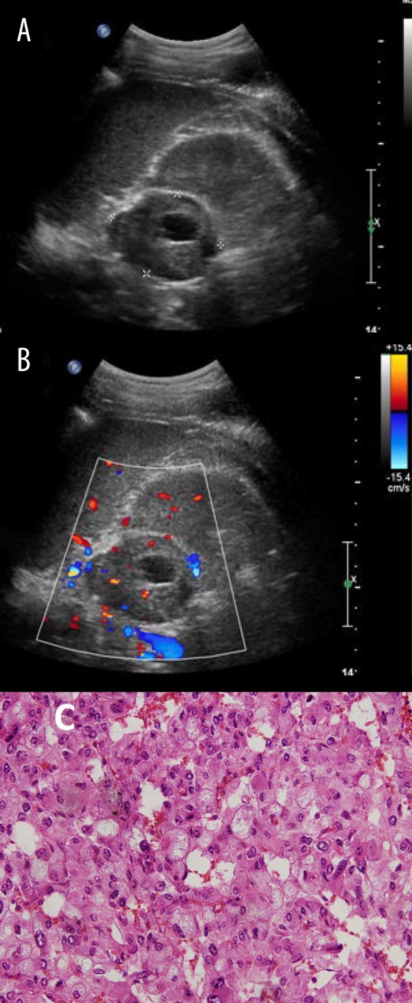
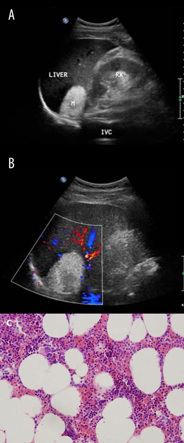
Results: There were 553 adenomas identified in the study, which constituted 60.70% of the lesions. There were 161 pheochromocytomas (17.67%), 49 myelolipomas (5.38%), 39 cysts (4.28%), 37 metastasis (4.06%), 35 ganglioneuromas (3.84%), 22 lymphomas (2.41%), and 15 cortical carcinomas (1.65%). The sensitivity, specificity, and accuracy of ultrasound-based diagnosis were 89%, 99%, and 93.9%, respectively. A positive predictive value of 90.9% and a negative predictive value of 94.2% were obtained in this study.
Conclusions: This large-sample study showed that ultrasound was a reliable method in differentiating benign 'leave-alone' lesions from suspicious lesions.
Figures
References
-
- Young WF., Jr Clinical practice. The incidentally discovered adrenal mass. N Engl J Med. 2007;356(6):601–10. - PubMed
-
- Grumbach MM, Biller BMK, Braunstein GD, et al. Management of the clinically inapparent adrenal mass (“incidentaloma”): NIH conference. Ann Intern Med. 2003;138(5):424–29. - PubMed
-
- Suzuki Y, Sasagawa, Suzuki H, et al. The role of ultrasonography in the detection of adrenal masses: comparison with computed tomography and magnetic resonance imaging. Int Urol Nephrol. 2001;32:303–6. - PubMed
-
- Lumachi F, Borsato S, Brandes AA, et al. Fine needle aspiration cytology of adrenal masses in noncancer patients: clinicoradiologic and histologic correlations in functioning and nonfunctioning tumors. Cancer. 2001;93:323–29. - PubMed
-
- Trojan J, Schwarz W, Sarrazin C, et al. Role of ultrasonography in the detection of small adrenal masses. Ultraschall Med. 2002;23:96–100. - PubMed
Publication types
MeSH terms
LinkOut - more resources
Full Text Sources
Other Literature Sources
Medical
