. 2015 Mar;172(5):1195-221.
doi: 10.1111/bph.12992. Epub 2015 Jan 13.

Synthesis and SAR studies of novel 6,7,8-substituted 4-substituted benzyloxyquinolin-2(1H)-one derivatives for anticancer activity

Affiliations

Synthesis and SAR studies of novel 6,7,8-substituted 4-substituted benzyloxyquinolin-2(1H)-one derivatives for anticancer activity

Yi-Fong Chen et al. Br J Pharmacol. 2015 Mar.

Abstract

Background and purpose: 4-Phenylquinolin-2(1H)-one (4-PQ) derivatives can induce cancer cell apoptosis. Additional new 4-PQ analogs were investigated as more effective, less toxic antitumour agents.

Experimental approach: Forty-five 6,7,8-substituted 4-substituted benzyloxyquinolin-2(1H)-one derivatives were synthesized. Antiproliferative activities were evaluated using a 3-(4,5-dimethylthiazol-2-yl)-2,5-diphenyltetrazoliun bromide assay and structure-activity relationship correlations were established. Compounds 9b, 9c, 9e and 11e were also evaluated against the National Cancer Institute-60 human cancer cell line panel. Hoechst 33258 and Annexin V-FITC/PI staining assays were used to detect apoptosis, while inhibition of microtubule polymerization was assayed by fluorescence microscopy. Effects on the cell cycle were assessed by flow cytometry and on apoptosis-related proteins (active caspase-3, -8 and -9, procaspase-3, -8, -9, PARP, Bid, Bcl-xL and Bcl-2) by Western blotting.

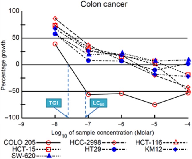
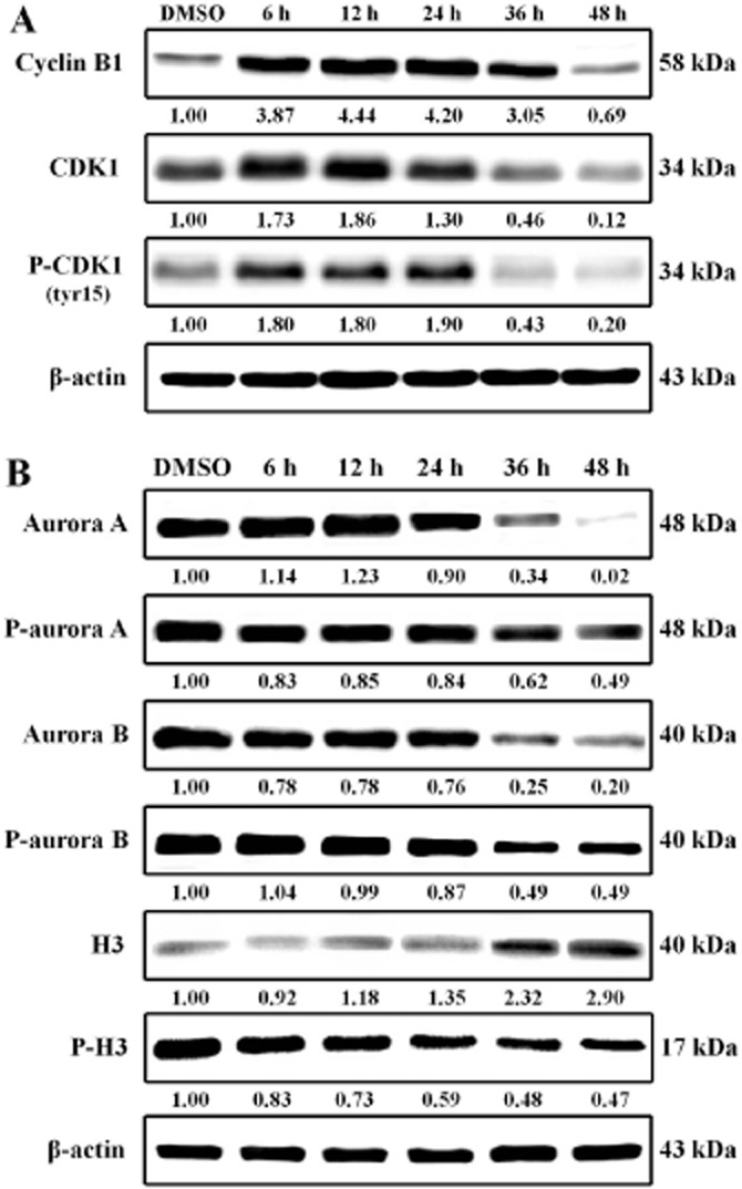
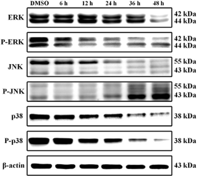
Key results: Nine 6,7,8-substituted 4-substituted benzyloxyquinolin-2(1H)-one derivatives (7e, 8e, 9b, 9c, 9e, 10c, 10e, 11c and 11e) displayed high potency against HL-60, Hep3B, H460, and COLO 205 cancer cells (IC₅₀ < 1 μM) without affecting Detroit 551 normal human cells (IC₅₀ > 50 μM). Particularly, compound 11e exhibited nanomolar potency against COLO 205 cancer cells. Mechanistic studies indicated that compound 11e disrupted microtubule assembly and induced G2/M arrest, polyploidy and apoptosis via the intrinsic and extrinsic signalling pathways. Activation of JNK could play a role in TRAIL-induced COLO 205 apoptosis.

Conclusion and implications: New quinolone derivatives were identified as potential pro-apoptotic agents. Compound 11e could be a promising lead compound for future antitumour agent development.


Figures

Figure 1
Figure 1
The structures of some anticancer agents and the general structure of the target compounds (7a–e ∼ 15a–e).
Figure 15
Figure 15
Scheme Reagents and conditions: (A) 130°C with PPA. (B) K2CO3/DMF, 80–90°C.
Figure 2
Figure 2
Alkylation of 4-hydroxyquinolin-2(1H)-ones. (A) Tautomerism of 4-hydroxyquinolin-2(1H)-one derivatives. (B) Key HMBC correlations (blue arrows) of 11e indicated alkylation at the 4-OH position.
Figure 3
Figure 3
Subpanel tumour cell lines selectivity ratios of selected compounds 9b, 9c, 9e and 11e.
Figure 4
Figure 4
Dose-response curves of compound 11e against colon cancer cell lines.
Figure 5
Figure 5
Compound 11e induced time-dependent apoptosis in COLO 205 cells. COLO 205 cells were treated with 50 nM of 11e for 0, 12, 24, 36 and 48 h. (A) Compound 11e induced morphological changes in COLO 205 cells. (B) Fluorescent images of Hoechst staining showing 11e-induced cell death. The black arrowhead indicates an apoptotic nucleus and the white arrowheads indicate multinucleate cells. (C) Apoptosis induced by compound 11e was confirmed using annexin V/PI staining and flow cytometry. The fraction of annexin V-positive COLO 205 cells was 5.5% prior to treatment and 7.6%, 12.7%, 20.9% and 21.8% after treatment with 11e for 12, 24, 36 and 48 h respectively. Scale bar = 20 μm.
Figure 6
Figure 6
Effects of 11e on the cytotoxicity of COLO 205 cells. (A) Chemical structure of 11e. (B) COLO 205 cells were exposed to different concentrations of 11e for 48 h. (C) COLO 205 cells were exposed to 0, 10, 25, 50, 75 and 100 nM 11e for 24, 48 and 72 h. Cell viability was assessed using the MTT assay. The data are presented as mean ± SEM of three independent experiments. Cells without treatment served as a control. *P < 0.001 versus control.
Figure 7
Figure 7
11e delays M phase progression and caused microtubule disassembly in cultured cells. (A) Flow cytometry analysis of cell cycle distribution in COLO 205 colon cancer cell line treated with 50 nM of 11e for 0, 6, 12, 24, 36 and 48 h. (B) The effect of 11e on the microtubule formation in COLO 205 cells. Cells were incubated with 0.1% DMSO, 50 nM 11e, 1 μM colchicine or 1 μM taxol for 24 h. Immunofluorescence for α-tubulin (green) and PI nuclear staining (red). Cells were visualized using confocal microscopy.
Figure 8
Figure 8
The docked binding mode of 11e is shown with the binding site of tubulin (PDB entry 1SA0). The figures were performed using PyMol. (A) The binding mode of DAMA-colchicine (red stick model) and tubulin. (B) The binding mode of 11e (yellow stick model) and tubulin. (C) DAMA-colchicine and 11e occupy similar binding space in tubulin (shown as surface of tubulin cavity). (D) The superimposition of DAMA-colchicine and 11e.
Figure 9
Figure 9
Compound 11e increased G2/M phase checkpoint protein expression. COLO 205 cells were treated with 50 nM 11e for the indicated time periods and lysed for protein extraction. Protein samples (40 μg protein per lane) were separated using 10% SDS-PAGE and subjected to immunoblotting with antibodies specific to cyclin B1, CDK1, phospho-CDK1 (A), aurora A, phospho-aurora A, aurora B, phospho-aurora B, H3, phospho-H3 (B) and β-actin (n = 3 independent experiments). β-Actin was used as a loading control.
Figure 10
Figure 10
Compound 11e induced caspase-3, caspase-8 and caspase-9 activity in COLO 205 cells. COLO 205 cells were treated with 50 nM 11e for the indicated times and lysed for protein extraction. Protein samples (40 μg protein per lane) were separated using 10% SDS-PAGE and subjected to immunoblotting with antibodies specific to caspase-9, caspase-8, caspase-3, PARP and β-actin (n = 3 independent experiments). β-Actin was used as a loading control.
Figure 11
Figure 11
Compound 11e induced the mitochondrial apoptosis pathway in COLO 205 cells. (A) Effects of 11e on mitochondrial membrane potential in COLO 205 cells. Cells (1 × 10⁶ cells·mL−1) were untreated or treated with 11e (50 nM, 6–48 h) to induce apoptosis. Cells were stained with JC-1 and analysed by flow cytometry. (B) COLO 205 cells were treated with 50 nM 11e for the indicated times and lysed for protein extraction. Protein samples (40 μg protein per lane) were separated using 10% SDS-PAGE and subjected to immunoblotting with antibodies specific to AIF, Endo G, Apaf-1, cytochrome c and β-actin (n = 3 independent experiments). (C) Compound 11e affected Bcl-2 family proteins in COLO 205 cells. COLO 205 cells were treated with 50 nM 11e for the indicated times and lysed for protein extraction. Protein samples (40 μg protein per lane) were separated using 10% SDS-PAGE and subjected to immunoblotting with antibodies specific to Bid, Bax, Bad, Bcl-xL, Bcl-2 and β-actin (n = 3 independent experiments). β-Actin was used as a loading control.
Figure 12
Figure 12
Compound 11e-induced death receptor apoptosis pathways in COLO 205 cells. COLO 205 cells were treated with 50 nM 11e for the indicated times and lysed for protein extraction. Protein samples (40 μg protein per lane) were separated using 10% SDS-PAGE and subjected to immunoblotting with antibodies specific to Fas, TNFR1, DR4, DR5 (A), FasL, TNF-α, TRAIL (B) and β-actin (n = 3 independent experiments). β-Actin was used as a loading control.
Figure 13
Figure 13
Expression of MAPKs in the 11e-treated COLO 205 cells. COLO 205 cells were treated with 50 nM 11e for the indicated times and lysed for protein extraction. Protein samples (40 μg protein per lane) were separated using 10% SDS-PAGE and subjected to immunoblotting with antibodies specific to ERK1/2, phospho-ERK1/2, JNK, phospho-JNK, p38, phospho-p38 and β-actin (n = 3 independent experiments). β-Actin was used as a loading control.
Figure 14
Figure 14
The signalling pathways of 11e-induced G2/M phase arrest and apoptosis in human colon cancer COLO 205 cells.

Similar articles

Cited by

References

    1. Abonia R, Insuasty D, Castillo J, Insuasty B, Quiroga J, Nogueras M, et al. Synthesis of novel quinoline-2-one based chalcones of potential anti-tumor activity. Eur J Med Chem. 2012;57:29–40. - PubMed
    1. Ahmed N, Brahmbhatt KG, Sabde S, Mitra D, Singh IP, Bhutani KK. Synthesis and anti-HIV activity of alkylated quinoline 2,4-diols. Bioorg Med Chem. 2010;18:2872–2879. - PubMed
    1. Ahmed N, Brahmbhatt KG, Singh IP, Bhutani KK. Efficient chemoselective alkylation of quinoline 2,4-diol derivatives in water. J Heterocycl Chem. 2011;48:237–240.
    1. Ahvale AB, Prokopcová H, Šefčovičová J, Steinschifter W, Täubl AE, Uray G, et al. 4-Cyano-6,7-dimethoxycarbostyrils with solvent- and pH-independent high fluorescence quantum yields and emission maxima. Eur J Org Chem. 2008;2008:563–571.
    1. Alexander SPH, Benson HE, Faccenda E, Pawson AJ, Sharman JL. Spedding M, et al. The Concise Guide to PHARMACOLOGY 2013/14: Enzymes. Br J Pharmacol. 2013a;170:1797–1867. - PMC - PubMed

Publication types

MeSH terms