. 2015 Jan 15;517(7534):377-80.
doi: 10.1038/nature13853. Epub 2014 Oct 26.

Control of plant stem cell function by conserved interacting transcriptional regulators

Affiliations

Control of plant stem cell function by conserved interacting transcriptional regulators

Yun Zhou et al. Nature. .

Abstract

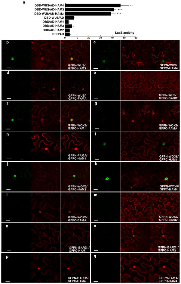
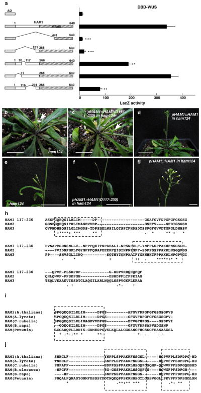
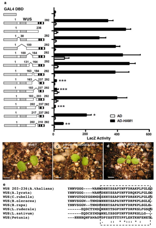
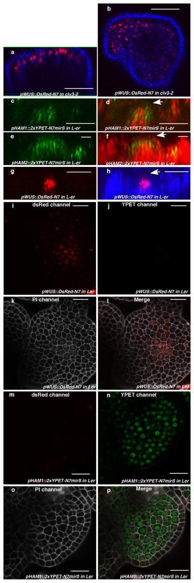
Plant stem cells in the shoot apical meristem (SAM) and root apical meristem are necessary for postembryonic development of aboveground tissues and roots, respectively, while secondary vascular stem cells sustain vascular development. WUSCHEL (WUS), a homeodomain transcription factor expressed in the rib meristem of the Arabidopsis SAM, is a key regulatory factor controlling SAM stem cell populations, and is thought to establish the shoot stem cell niche through a feedback circuit involving the CLAVATA3 (CLV3) peptide signalling pathway. WUSCHEL-RELATED HOMEOBOX 5 (WOX5), which is specifically expressed in the root quiescent centre, defines quiescent centre identity and functions interchangeably with WUS in the control of shoot and root stem cell niches. WOX4, expressed in Arabidopsis procambial cells, defines the vascular stem cell niche. WUS/WOX family proteins are evolutionarily and functionally conserved throughout the plant kingdom and emerge as key actors in the specification and maintenance of stem cells within all meristems. However, the nature of the genetic regime in stem cell niches that centre on WOX gene function has been elusive, and molecular links underlying conserved WUS/WOX function in stem cell niches remain unknown. Here we demonstrate that the Arabidopsis HAIRY MERISTEM (HAM) family of transcription regulators act as conserved interacting cofactors with WUS/WOX proteins. HAM and WUS share common targets in vivo and their physical interaction is important in driving downstream transcriptional programs and in promoting shoot stem cell proliferation. Differences in the overlapping expression patterns of WOX and HAM family members underlie the formation of diverse stem cell niche locations, and the HAM family is essential for all of these stem cell niches. These findings establish a new framework for the control of stem cell production during plant development.


Conflict of interest statement

The authors declare no competing financial interests.

Figures

Extended Data Figure 1
Interaction between WUS/WOX and HAM family transcriptional regulators. (a) LacZ activity in yeast-two-hybrid assays testing interactions between WUS and HAM2, HAM3 or HAM4. Error bar = mean ± sem (n=3biological replicates). **, P<0.01; ***, P<0.001(two-tailed t-test, compared to DBD-WUS/AD). (bo) Bimolecular fluorescence complementation (BiFC) analyses in tobacco transient assays with HAM and WOX family genes. Tobacco was co-transformed with GFPn-WUS and GFPc-HAM3 (b), or GFPn-WUS and GFPc-HAM4 (c), or GFPn-WUS and GFPc-FAMA (d), or GFPn-WUS and GFPc-BARD1 (e), or GFPn-WOX4 and GFPc-HAM1 (f), or GFPn-WOX4 and GFPc-FAMA (g), or GFPn-FAMA and GFPc-HAM1 (h), or GFPn-WOX5 and GFPc-HAM1 (i), or GFPn-WOX5 and GFPc-HAM2 (j), or GFPn-WOX5 and GFPc-HAM4 (k), or GFPn-WOX5 and GFPc-FAMA (l), or GFPn-WOX5 and GFPc-BARD1 (m), or GFPn-BARD1 and GFPc-HAM1 (n), or GFPn-BARD1 and GFPc-HAM2 (o), or GFPn-BARD1 and GFPc-HAM4 (p), or GFPn-FAMA and GFPc-HAM4 (q). BARD1 and FAMA proteins are both included as negative controls. Left panel: GFP channel; middle panel: propidium iodide (PI) staining channel; right panel: merged channels. Scale bar = 20 μm.
Extended Data Figure 2
An N-terminal region of HAM1 is important for WUS-HAM1 interactionand is essential for HAM1 function in stem cell maintenance. (a) Yeast two-hybrid assay of interactions between WUS and various deleted derivatives of HAM1. Deleting amino acids 117 to 230(D117-230) from HAM1 compromised the WUS-HAM1 interaction. Left panel: box diagrams of the HAM1 derivatives. Shaded boxes indicate the GRAS domains. Numbers indicate amino acid residues. Error bar = mean ± sem (n=3biological replicates). *, P<0.05; **, P<0.01; ***, P<0.001(two-tailed t-test, compared to AD-HAM1 full-length). (bg) The complementation of the ham1;2;4 triple mutant requires amino acids 117-230. The early termination phenotype of ham1;2;4(b, e) was not complemented by HAM1 (D117-230) driven by a HAM1 promoter and 3′UTR (c, f), but was fully complemented by wild type HAM1(d, g). Arrows in (b, c) indicate the early-terminated inflorescences. (hj) Amino acid sequence alignment of the HAM1 N-terminal domains (117–230) using Clustal Omega. (h) Sequence alignment of the N-terminal domains among three Arabidopsis HAM members. (i) Sequence alignment of partial N-terminal domains in HAM from A.thaliana, A.lyrata, C.rubella, B.rapa, and Petunia. (j) Sequence alignment of partial HAM1 N-terminal domains in HAM from A.thaliana, A.lyrata, C.rubella, B.oleracea, B.rapa, and Petunia. Asterisks: same amino acids; dots: similar amino acids. The conserved regions are boxed. Bars = 10 mm in (b, c), and (g), 40 mm in (d), 20 mm in (e, f).
Extended Data Figure 3
A C-terminal region of WUS is important for WUS-HAM1 interaction and is essential for WUS function in stem cell maintenance. (a) Yeast-two-hybrid assay of interactions between HAM1 and various deleted WUS derivatives. Deleting amino acids 203 to 236 (D203-236) from WUS greatly compromised the WUS-HAM1 interaction. Left panel: box diagrams of the deleted WUS derivatives; shaded boxes: the homeodomain; three black boxes: the acidic domains, the WUS box and the EAR motif, respectively. Numbers indicate amino acid residues. Error bar = mean ± sem (n=3biological replicates).*, P<0.05; **, P<0.01; ***, P<0.001(two-tailed t-test, compared to DBD-WUS full-length). (bd) WUS function requires the same region that is important for WUS-HAM1 interaction. The terminated shoot meristem phenotype of wus-1 (b) was not complemented by WUS (D203-236) driven by WUS promoter and 3′UTR (c), and was fully complemented by the wild type WUS (d). (e) Amino acid sequence alignment of C-terminal regionsof WUS from Arabidopsis thaliana, A.lyrata, Capsella rubella, Brassica oleracea, B.rapa, Lepidium ruderale, L.sativum, and Petunia, using Clustal Omega. Asterisks: same amino acids; dots: similar amino acids. The conserved regions are boxed. Bars = 2 mm in (bd).
Extended Data Figure 4
Genetic interaction between WUS and HAM family members. (a, b) The secondary inflorescence meristems initiated from axillary meristems in wus-7; ham1;2 homozygotes with ham3/+ terminate prematurely. (c)wus-7; ham1;2 homozygotes with ham4/+ display early termination of the main inflorescence meristem and lack of carpels in flowers (indicated by arrow). (df) WUS and HAM family members interact genetically in a dose-dependent manner. wus-7 (d) formed functional shoot apices and normal stature, but wus-7; ham1/+; ham2/+; ham3/+ (e) enhanced the wus-7 phenotype, and wus-7; ham1/+; ham2; ham3 (f) showed stronger enhancement, with reduced flower numbers and plant stature, and elongated vegetative stage, resembling a wus strong allele. Plants are 36 days after germination. (gh) Down-regulation of HAM1, HAM2 and HAM3 in ham4 shoot meristems leads to an early termination phenotype. Compared to wild type (Col) (g), pWUS::mir171 in ham4 (h) showed terminated vegetative meristems. (il) WUS is required for functions of HAM1, HAM2 and HAM3. At 11 days after germination, compared to Ler wild type (i) and ham1;2;3 (k) which formed functional vegetative meristem and leaf primordia, wus-1; ham1;2;3 (l) displays terminated vegetative meristems similar to wus-1 (j). Bars = 2 mm.
Extended Data Figure 5
Expression of HAM1, HAM2 and WUS in the SAMs. (ab) WUS expression in clv3-2. Orthogonal (a) andtop (b) views of pWUS::DsRed-N7 expression (red)and chlorophyll autofluorescence (blue) in the same clv3-2 inflorescence meristem. (ch) Comparison between expression patterns of HAM1, HAM2 and WUS in vegetative meristems.(c) Orthogonal view of pHAM1::2xYPET-N7mirS expression (green) in Ler vegetative meristem. (d) Orthogonal view of pHAM1::2xYPET-N7mirS expression (green) together with chlorophyll autofluorescence (red) in the same vegetative meristem shown in (c), indicating that HAM1 is expressed in the rib meristem. (e) Orthogonal view of pHAM2::2xYPET-N7mirS expression (green) in Ler vegetative meristem. (f) Orthogonal view of pHAM2::2xYPET-N7mirS expression (green) together with chlorophyll autofluorescence (red) in the same vegetative meristem shown in (e), indicating that HAM2 is highly expressed in the rib meristem. (g) Orthogonal view of pWUS::DsRed-N7 expression (red) in Ler vegetative meristem. (h) Orthogonal view of pWUS::DsRed-N7 expression (red) together with chlorophyll autofluorescence (blue) in the same vegetative meristem shown in (g), indicating that WUS is expressed in the rib meristem. Arrows indicate the positions of L1 cell layer. (ip) Control images confirming the specificity of confocal spectral settings for Fig. 3(el). The SAMs from pWUS::DsRed-N7 line (il) or pHAM1::2xYPET-N7mirS line (mp) were imaged from the same three separated channels used in Fig. 3 (el). There is no spectral bleed-through of YPET signal into the ds Red channel (m), nor ds Red signal into the YPET channel (j). (i, m)ds Red channel (red); (j, n) YPET channel (green); (k, o) PI staining channel (gray); (l, p) merged all three channels. Bars = 50μm in (ad, gh), 20 μm in (ef, ip).
Extended Data Figure 6
pHAM2::YFP-HAM2 (pHAM2::YPET-HAM2)complemented the ham1,2,4 mutant and was expressed in the center of SAMs. The early termination phenotype ofham1;2;4(a, b) was completely complemented by YPET-HAM2 driven by the HAM2 promoter and 3′UTR (c), indicating the promoter used for HAM2 transcriptional and translational reporters are functional and the fusion protein (YPET-HAM2) is also functional in vivo. Arrows in (a, b) indicate early-terminated apices. Bars = 10 mm (ac). (de) Different Z sections from the same SAM in ham1,2,4 [pHAM2::YPET-HAM2] plant for Fig. 3 (mn) show expression of pHAM2::YPET-HAM2 translationalmarker (green) in L2 (d) and L3 (e), together with PI as counter stain (Red). Bars = 20 μm (de). (f) Immunoblot with anti-GFP antibody validates the presence of YFP-HAM2 (YPET-HAM2) in both nuclear lysate and nuclear proteins immunoprecipitated with GFP-Trap from ham1,2,4 [pHAM2::YFP-HAM2] line used in ChIP experiment (Fig. 2, n and o).
Extended Data Figure 7
Expression patterns of HAMgenesin comparison to WOX4. (a) pHAM4::2xYPET-N7 (green, arrow indicated) is expressed in procambium cells of the first leaf. (b) pHAM4::2xYPET-N7 (green, arrow indicated) is expressed in vasculature in the 7-day-old hypocotyl. (ch) Comparison of pHAM4::2xYPET-N7 (green, arrow indicated) and pWOX4::YFP (green, arrow indicated) expression patterns in vasculature cells in the 7-d-old leaf petiole (c–d),20-d-old leaf petiole (e–f) and 7-d-old root (g–h). (i) Orthogonal view of pHAM4::2xYPET-N7 (green, arrow indicated) expression in flower vasculature. (j) Procambium-specific expression of pHAM4::2xYPET-N7 in stems from 1cm bolting plants. (k–l) Procambium-specific expression of pHAM3::2xYPET-N7mirS (k) and pHAM1::2xYPET-N7mirS (l) in transverse sections of stems from 1 cm bolting plants. Red in (a–f, i–l) represents chlorophyll autofluorescence, and represents PI staining in (g–h). Bars = 50μm in (a, h–i, k–l); 100μm in (b–g, j).
Extended Data Figure 8
Expression patterns of HAM2 transcriptional and translational reporters in root meristems. (a–i) Complete stacks of confocal sections through the root tip demonstrate that pHAM2::2xYPET-N7mirS (green) is expressed in the QC cells (arrow indicated) and cells above the QC within the root meristem. (j–o) Expression patterns of HAM2 translational reporters in ham1,2,4 root meristems. Complete stacks of confocal sections through the root tip demonstrate that pHAM2:YPET-HAM2 (green) is present in the QC cells (arrow indicated) and the cells above the QC within the root meristem in the ham1,2,4 mutant. In all figures cellular outlines were stained with PI (red). Bars = 20μm (a–i), 50μm (j–o).
Extended Data Figure 9
HAM family regulates various stem cell niches. (a–d) Growth arrest of ham1;2;3;4 at the seedling stage. Imaging of Ler wild type (WT) (a) and homozygous ham1;2;3;4 (b) seedlings at 7 days after germination (DAG). Imaging of WT (c) and homozygous ham1;2;3;4 (d) (arrow indicated) seedlings at 26 DAG. (e–f) Transverse section of leaves from WT (e) and ham1;2;3;4 (f) at 7 DAG. Arrow in (f) indicates undifferentiated/undetermined cell mass. (g–h) Confocal imaging of root meristem from WT (g) and ham1;2;3;4 (h) seedlings at 7 DAG. ham1;2;3;4 displayed enlarged cells with abnormal shapes at the QC (arrow indicated) and columella stem cell (CSC) positions. Cellular outlines (g–h) were visualized with PI staining (white). (i–l) mPS-PI stains indicate that HAM genes regulate root cell differentiation. Some CSCs (arrow indicated) undergo differentiation with starch accumulated and stained in homozygous ham1;2;3 (j, l), but none of them can be stained in L-er wild type (i, k). Asterisks mark the QC cells. Bars= 5mm (c–d), 1 mm in (a–b, e–f), 20 μm in (g–l).
Extended Data Figure 10
Interaction between WOX and HAM homologs from tomato (Solanum lycopersicum). Bimolecular fluorescence complementation (BiFC) analyses in tobacco transient assays demonstrated that tomato WUS (Gene ID: 543793) physically interacted with a putative tomato HAM homolog (LEFL2052P11) (a) identified based on its sequence homology to HAM from Arabidopsis and Petunia (f), and tomato WOX4 (Gene ID: 100301933) physically interacted with the putative tomato HAM homolog (b). BARD1 protein is included as a negative control (c–e). Left panel: GFP channel; middle panel: PI staining channel; right panel: merged channels. Scale bar = 20 μm.(f) Amino acid sequence alignment of a putative tomato HAM, Arabidopsis HAM1 and petunia HAM using Clustal Omega. Asterisks: same amino acids; dots: similar amino acids.
Figure 1
Figure 1. WUS/WOX and HAM family proteins physically interact
(a) LacZ activity of yeast-two-hybrid assays. Error bar = mean ± sem (n=3biological replicates). ***, P<0.001(two-tailed t-test). (bd) Bimolecular fluorescence complementation in tobacco. Panels (left to right): GFP; propidium iodide (PI) staining; merged channels. Scale bar = 20μm.(e) SDS-PAGE of input recombinant proteins stained by Coomassie Blue (left), and pull down of His6-tagged HAM proteins through GST-tagged WUS/WOX proteins detected by immunoblotting with anti-His antibody (right). Asterisk: HAM1-His6band. (fj) Co-immunoprecipitation of WUS-GFP and FLAG-HAM1 (f), WUS-GFP and FLAG-HAM2 (g), GFP-WOX4 and FLAG-HAM4(h), WOX5-GFP and FLAG-HAM2(i) (see Methods).
Figure 2
Figure 2. WUS and HAM family genes cooperatively control the shoot stem cell niche and co-regulate a common gene set
Shoot apices (a–d) (arrows) and inflorescence structures(e–h)of plants in indicated genotypes. Bars = 2 mm in (a–h).(i) RT-PCR quantification of WUS and HAM target gene expression in indicated genotypes. Error bar = mean ± sem(n=3 biological replicates). (j–m) LUC/REN activity in tobacco cells co-transformed with different reporter constructs (structured above each graph) and indicated effectors(see Methods). Min35S: 60 base pair 35S minimum element, REN: Renilla luciferase, LUC: firefly luciferase, LB/RB: T-DNA left or right border. Error bar = mean ± sem(n=3biological replicates). (n–o) Chromatin Immunoprecipitation of HAM2 protein with TPL or GRP23 chromatin regions, with ampliconlocations (Bars with numbers) diagrammed above each graph. The ChIP experiments were repeated three times using independent biological replicates with similar results, and one representative data set is shown.*, P<0.05; **, P<0.01; ***, P<0.001(two-tailed t-test) in (i–o).
Figure 3
Figure 3. HAM and WUS/WOX expression domains overlap
Expression of (a) pHAM1::2xYPET-N7microRNAsensitive marker (pHAM1::2xYPET-N7mirS) (green), (b) pHAM2::2xYPET-N7mirS (green), (c) pWUS::DsRed-N7 (red) in L-er inflorescence meristem (IM), and (d) pHAM1::2xYPET-N7mirS (green) marker in a clv3-2 IM. Orthogonal (upper panels) and transverse section (lower panels) views of the same plant are shown. (el) Overlapping expression patterns of pWUS::DsRed-N7 with pHAM1::2xYPET-N7mirS or pHAM2::2xYPET-N7mirS in the same shoot meristems (see Methods). Panels (from left to right): dsRed (red); YPET (green); PI (gray); merged channels. (mn) Expression of pHAM2::YPET-HAM2 translation almarker (green) in L1 (m) and L3 (n) of the same ham1,2,4 SAM.(ot) Overlapping expression patterns of pHAM4::2xYPET-N7and pWOX4::YFP (green, arrows) in the provascular and procambium cells in cotyledons (op), seedlings (qr), and stem transverse sections (st). PIcounter stain: red (ab, d, mn), green (c), gray (gh, kl). Chlorophyll autofluorescence: red (ot). Bars = 50 μm in (d, st); 200 μm in (o); 100 μm in (pr); 20 μm in (ac, en).
Figure 4
Figure 4. HAM family members are essential for various plant stem cell activities
Scanning electron microscopic imaging of WT (a) and ham1, ham2, ham3, ham4 (bd) seedlings (26 DAG). Arrow: ham1;2;3;4lacking a functional SAM.(ef) Transverse sections of WT and ham1;2;3;4hypocotyls (7 DAG). Bars = 1 mm in (ac, ef), 50 μm in (d).

Comment in

References

    1. Meyerowitz EM. Genetic control of cell division patterns in developing plants. Cell. 1997;88:299–308. - PubMed
    1. Sablowski R. The dynamic plant stem cell niches. Current opinion in plant biology. 2007;10:639–644. - PubMed
    1. Miyashima S, Sebastian J, Lee JY, Helariutta Y. Stem cell function during plant vascular development. The EMBO journal. 2013;32:178–193. - PMC - PubMed
    1. Dinneny JR, Benfey PN. Plant stem cell niches: standing the test of time. Cell. 2008;132:553–557. - PubMed
    1. Laux T, Mayer KF, Berger J, Jurgens G. The WUSCHEL gene is required for shoot and floral meristem integrity in Arabidopsis. Development. 1996;122:87–96. - PubMed

Publication types