Review
. 2014 Dec;76(6):769-83.
doi: 10.1002/ana.24291. Epub 2014 Nov 7.

Neuroimaging biomarkers for Parkinson disease: facts and fantasy

Affiliations
Review

Neuroimaging biomarkers for Parkinson disease: facts and fantasy

Joel S Perlmutter et al. Ann Neurol. 2014 Dec.

Abstract

In this grand rounds, we focus on development, validation, and application of neuroimaging biomarkers for Parkinson disease (PD). We cover whether such biomarkers can be used to identify presymptomatic individuals (probably yes), provide a measure of PD severity (in a limited fashion, but frequently done poorly), investigate pathophysiology of parkinsonian disorders (yes, if done carefully), play a role in differential diagnosis of parkinsonism (not well), and investigate pathology underlying cognitive impairment (yes, in conjunction with postmortem data). Along the way, we clarify several issues about definitions of biomarkers and surrogate endpoints. The goal of this lecture is to provide a basis for interpreting current literature and newly proposed clinical tools in PD. In the end, one should be able to critically distinguish fact from fantasy.


Figures

Figure 1
Figure 1
Depiction of presynaptic and postsynaptic terminals in a nigrostriatal dopaminergic synapse. Tyr = tyrosine; Dopa = dihydroxyphenylalanine; DA = dopamine; D1 = dopamine D1-like receptor; D2 = dopamine D2-like receptor; [11C]DTBZ = [11C]dihydrotetrabenazine; [18F]FD = 6-[18F]fluorodopa; [11C]CFT = 2-beta-[11C]carbomethoxy-3-beta-4-fluorophenyltropane, DAT = dopamine active transporter; VMAT2 = vesicular monoamine transporter 2; AC = adenylyl cyclase.
Figure 2
Figure 2
MPTP induced nigrostriatal injury in non-human primates detected by PET. PET images in the same non-human primate prior to unilateral right intracarotid administration of MPTP (left column) and 8 weeks after MPTP administration (right column). Each row represents PET imaging performed with a different presynaptic radiopharmaceutical: [11C]DTBZ = [11C]dihydrotetrabenazine , [18F]FD = 6-[18F]fluorodopa, and [11C]CFT = 2-beta-[11C]carbomethoxy-3-beta-4-fluorophenyltropane. Note unilateral reduction (right brain) of radioactivity in the caudate and putamen following ipsilateral MPTP injection. Right brain is represented on the right side of the image.
Figure 3
Figure 3
Linear correlations of the right-to-left ratios (R/L) of striatal FD uptake constants with R/L ratios of TH activity (as measured using optical densitometry) following unilateral MPTP administration. The lines are the best fit and 90% confidence interval. Note, these data are bimodally distributed and invalidate the fit and correlation comparing in vitro cell counts with in vivo SPECT measures. The two clusters may account for those strongly affected and those relatively unaffected by an intervention. Figure adapted from Pate et al, 1993.
Figure 4
Figure 4
Regression analysis comparing in vivo DAT SPECT imaging with in vitro nigrostriatal measures. A pin-hole high resolution SPECT scanner was used to image in vivo striatal [123I]FP-CIT uptake in rats treated unilaterally with 6-hydoxydopamine (6-OHDA, which destroys dopaminergic neurons). Regression analysis of A) in vitro measures of striatal DA concentration and striatal [123I]FP-CIT binding ratio, and B) unbiased stereologic in vitro counts of nigral TH+ cell number and striatal [123I]FP-CIT binding ratio. Note, these data are bimodally distributed and invalidate the regression analysis comparing the in vitro cell counts with the in vivo SPECT measures. Values for lesioned and contralateral sham-lesioned hemisphere are shown as black and white spots, respectively (n=26). Figure and legend adapted from Alvarez-Fischer et al, 2007.
Figure 5
Figure 5
PET correlates with nigrostriatal damage and parkinsonism following unilateral intracarotid MPTP in non-human primates. A) Digital subtraction lateral projection cerebral angiography in a non-human primate demonstrating the location of the unilateral intra-carotid catheter tip just prior to injection of MPTP. B) Plot demonstrating the behavioral response in different animals given varying doses of MPTP (y axis = degree of parkinsonism) over 2 months (x axis = time) following unilateral intra-carotid MPTP administration. Error bars represent standard deviations (N=15). C) Parkinsonism score at 2 months versus percent of residual dopaminergic cell counts and D) dopamine content in the striatum (n=14). Data from monkeys with residual nigral cell counts of less than 50% are depicted by circles (n=5). Squares represent data from monkeys with residual nigral cell counts of 50% or more (n=9). Figure and legend adapted from Tabbal et al., 2012.
Figure 6
Figure 6
Relationship between Striatal dopamine and and substantia nigra TH+ cell counts: Percent of residual dopaminergic cell counts (measured with unbiased stereology) in injected nigra (x-axis) versus percent residual dopamine (measured with HPLC) in the striatum (y-axis, n=14). Data from non-human primates with residual nigral cell counts of less than 50% are depicted in circles (n=5). Squares represent data from primates with residual nigral cell counts of 50% or more (n=9). Figure and legend adapted from Tabbal et al, 2012.
Figure 7
Figure 7
PET measures correlated with striatal dopamine. Relationship of in vitro measures of striatal dopamine (% control side) compared to in vivo PET measures of striatal A) FD KOCC, B) CFT and C) DTBZ BPND (% control side). Correlation is significant with or without the clustered data points in the lower left corner of each graph. Figure and legend adapted from Karimi et al, 2013.
Figure 8
Figure 8
PET measures have flooring effect in relation to nigral cell counts. Relationship between in vivo PET measures of FD KOCC ratio, CFT and DTBZ BPND ratio with in vitro measures of nigral dopaminergic neurons. All of the striatal PET measures approached zero once nigral cell loss reached 50%. Figure and legend adapted from Karimi et al, 2013.
Figure 9
Figure 9
In vivo PET measures of VMAT2 and DAT correlated with in vitro measures. The relationship between A) VMAT2 or B) DAT Bmax and residual substantia nigra pars compacta tyrosine hydroxylase immunoreactive neurons. The value for each non-human primate was expressed as the ratio of the injected side to the control (n = 13). Thus, these relationships do not depend upon specific molecular imaging radiopharmaceutical but rather reflect underlying pathophysiology. Figure and legend adapted from Tian et al, 2012.
Figure 10
Figure 10
Substantia nigra (SN) PET imaging correlated with nigral cell counts. The relationship between A) CFT and B) DTBZ injected/control side non-displaceable binding potential (or influx constant [Kocc]) in the SN and injected/control side ratio of TH-positive neurons in the SN. Each data point represents 1 non-human primate, and the trend lines are the linear fits of the data. CFT and DTBZ exhibited a tight linear correlation with residual cells in the SN. Midbrain based PET measures correlate with nigral cell counts whereas striatal terminal field measures do not. Figure and legend adapted from Brown et al, 2012.
Figure 11
Figure 11
FD PET reduced striatal uptake endophenotype. A) [18F]FDOPA PET in early PD with reduced uptake in posterior putamen compared to B) an age and gender matched control. C) Ki in control participants (with [n=48] and without [n=24] PD) and Amish (n=11). Forty-seven (98%) of 48 PD controls and four (36%) Amish met PET criteria for PD (Ki for one of the posterior putamen more than three standard deviations below the normal mean) despite clinical features not convincing for or against PD. By categorizing 4 of these as “affected” based upon FD PET phenotype the LOD score improved for the entire linkage analysis. Figure and legend adapted from Racette et al, 2006.
Figure 12
Figure 12
FD PET reduced uptake in nigrostriatal pathway in asymptomatic welders differs from PD. FDOPA PET composite images of decay-corrected counts from 24 to 94 minutes from a representative A) control. B) welder, and C) subject with idiopathic Parkinson disease (IPD) normalized to the reference region. FDOPA uptake is reduced in the caudate region of the welder in comparison to the control subject while the posterior putamen is the most affected region in the subject with IPD. Data previously presented in Criswell et al, 2011.
Figure 13
Figure 13
PiB PET does not distinguish subtypes of cognitive impairment in PD. Mean cortical binding potentials (MCBPs) for participant groups that include CTRL = healthy control with a Clinical Demetia Rating (CDR =0; n=48; 6% PiB+), CTRL-CI = control with cognitive impairment (CDR = 0.5; n=2; 100% PiB+), PD-nl = Parkinson disease, no cognitive impairment (CDR = 0; n=66; 9% PiB+), PD-CI = Parkinson disease, cognitive impairment (CDR = 0.5; n = 67; 15% PiB+), PD-dem = Parkinson disease with dementia (CDR = 1 or more; n = 24; 21% PiB+). Each point represents an individual participant (N = 207). The horizontal scale crossing the Y-axis at 0.18 is the recommended cut-off for PIB+. Figure and legend updated from data presented by Foster et al, 2010.
Figure 14
Figure 14
PiB scans target abnormal Aβ deposition, but not α-synuclein. [11C]-PIB PET images for A) Case 1 and B) Case 2 demonstrates increased signal in multiple cortical areas, including orbitofrontal and prefrontal cortex, precuneus, and temporal lobes. C) Case 3 and D) a control participant have minimal PIB signal in cortical areas. PIB retention in white matter areas is likely due to nonspecific binding. Data previously presented in Burack et al, 2010.
Figure 15
Figure 15
Distribution of principal component analysis component weights differ between PD and Alzheimer patients. Scatterplot represents the component weights of the primary principal components (PC1 and PC2) for the direct comparison of study participants with PD and Alzheimer disease. Note, that this distinction exists when limiting this analysis to those PD and AD patients with comparable degrees of cognitive impairment. Data previously presented in Campbell et al, 2011.
Figure 16
Figure 16
Comparison of survival in demented PD patients with postmortem findings of only synucleinopathy versus synucleinopathy plus abnormal Aβ deposition. Kaplan-Meier survival curves show A) the percentage of survival with respect to time from the onset of PD and B) time from the PD onset to dementia. Data previously presented in Kotzbauer et al, 2012.

References

    1. Bagchi DP, Yu L, Perlmutter JS, et al. Binding of the radioligand SIL23 to alpha-synuclein fibrils in Parkinson disease brain tissue establishes feasibility and screening approaches for developing a Parkinson disease imaging agent. PLoS One. 2013;8:e55031. - PMC - PubMed
    1. Brooks DJ, Frey KA, Marek KL, et al. Assessment of neuroimaging techniques as biomarkers of the progression of Parkinson's disease. Exp Neurol. 2003;184(Suppl 1):S68–S79. - PubMed
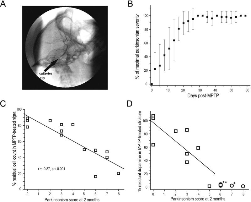
    1. Perlmutter JS, Tempel LW, Black KJ, et al. MPTP induces dystonia and parkinsonism: Clues to the pathophysiology of dystonia. Neurology. 1997;49:1432–1438. - PubMed
    1. Burn DJ, Mark MH, Playford ED, et al. Parkinson's disease in twins studied with 18F-dopa and positron emission tomography. Neurology. 1992;42:1894–1900. - PubMed
    1. Nagai Y, Obayashi S, Ando K, et al. Progressive changes of pre- and post-synaptic dopaminergic biomarkers in conscious MPTP-treated cynomolgus monkeys measured by positron emission tomography. Synapse. 2007;61:809–819. - PubMed

Publication types