

Progressive polyradiculoneuropathy due to intraneural oxalate deposition in type 1 primary hyperoxaluria
- PMID: 25363903
- PMCID: PMC4577279
- DOI: 10.1002/mus.24495
Progressive polyradiculoneuropathy due to intraneural oxalate deposition in type 1 primary hyperoxaluria
Abstract
Introduction: A 24-year-old man with primary hyperoxaluria type 1 (PH1) presented with a rapidly progressive axonal and demyelinating sensorimotor polyradiculoneuropathy shortly after the onset of end-stage renal disease. His plasma oxalate level was markedly elevated at 107 µmol/L (normal<1.8 µmol/L).
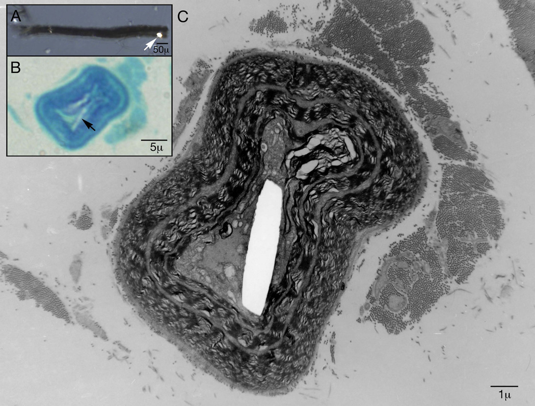
Methods: A sural nerve biopsy was performed. Teased fiber and paraffin and epoxy sections were done and morphometric procedures were performed on this sample and on an archived sample from a 22-year-old man as an age- and gender-matched control. Embedded teased fiber electron microscopy was also performed.
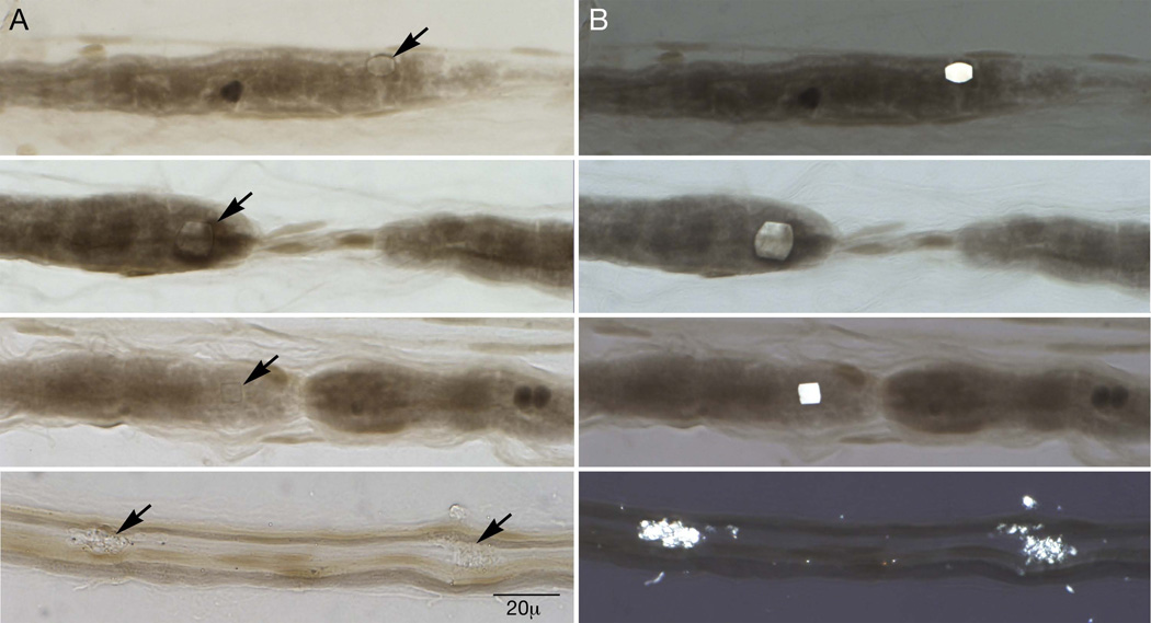
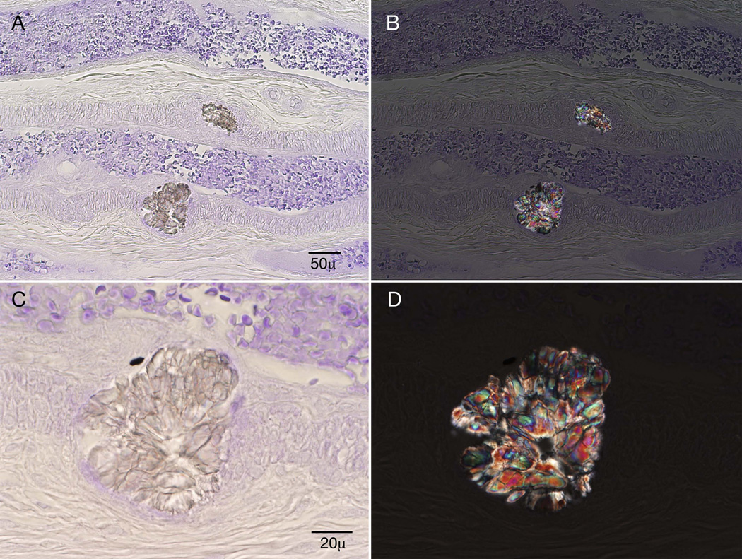
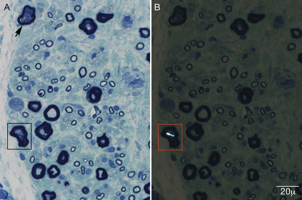
Results: The biopsy revealed secondary demyelination and axonal degeneration. Under polarized light, multiple bright hexagonal, rectangular, and starburst inclusions, typical of calcium oxalate monohydrate crystals, were seen.
Conclusions: The proposed mechanisms of nerve damage include disruption of axonal transport due to crystal deposition, toxic effect of oxalate, or nerve ischemia related to vessel occlusion from oxalate crystal deposition.
Keywords: crystalline neuropathy; nerve pathology; peripheral neuropathy; primary hyperoxaluria type 1; renal failure.
© 2014 Wiley Periodicals, Inc.
Figures
References
-
- Bilbao JM, Berry H, Merotta J, Ross RC. Peripheral neuropathy in oxalosis. A case report with electron microscopic observations. Le Journal Canadien Des Sciences Neurologiques. 1976;3(1):63–67. - PubMed
-
- Daudon M, Jungers P, Bazin D. Peculiar morphology of stones in primary hyperoxaluria. New England Journal of Medicine. 2008;359(1):100–102. - PubMed
-
- Illies F, Bonzel KE, Wingen AM, Latta K, Hoyer PF. Clearance and removal of oxalate in children on intensified dialysis for primary hyperoxaluria type 1. Kidney Int. 2006 Nov;70(9):1642–1648. - PubMed
Publication types
MeSH terms
Substances
Supplementary concepts
Grants and funding
LinkOut - more resources
Full Text Sources
Other Literature Sources
