

Oral mucocele: A clinical and histopathological study
- PMID: 25364184
- PMCID: PMC4211243
- DOI: 10.4103/0973-029X.141370
Oral mucocele: A clinical and histopathological study
Abstract
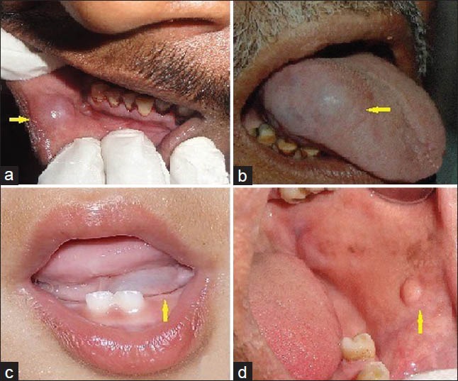
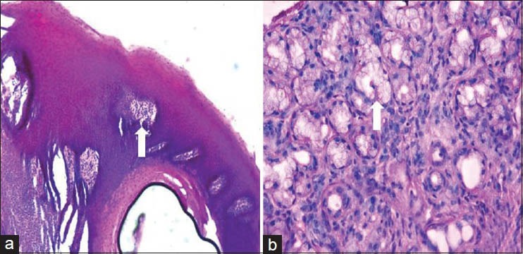
Background: Oral mucocele is the most common benign minor (accessory) salivary gland lesion, caused due to mechanical trauma to the excretory duct of the gland. Clinically they are characterized by single or multiple, soft, fluctuant nodule, ranging from the normal color of the oral mucosa to deep blue. It affects at any age and is equally present in both sexes with highest incidence in second decade of life. They are classified as extravasation or retention type.
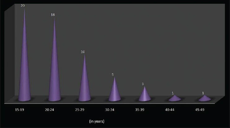
Objectives: To analyze the data between 2010 and 2011 of, clinically and histopathologically diagnosed 58 oral mucoceles for age, gender, type, site, color, cause, symptoms and dimension.
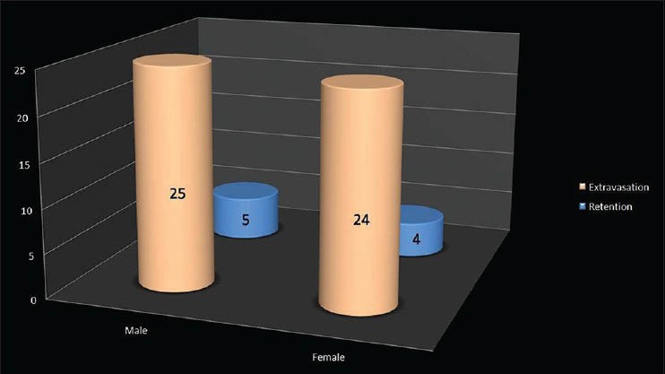
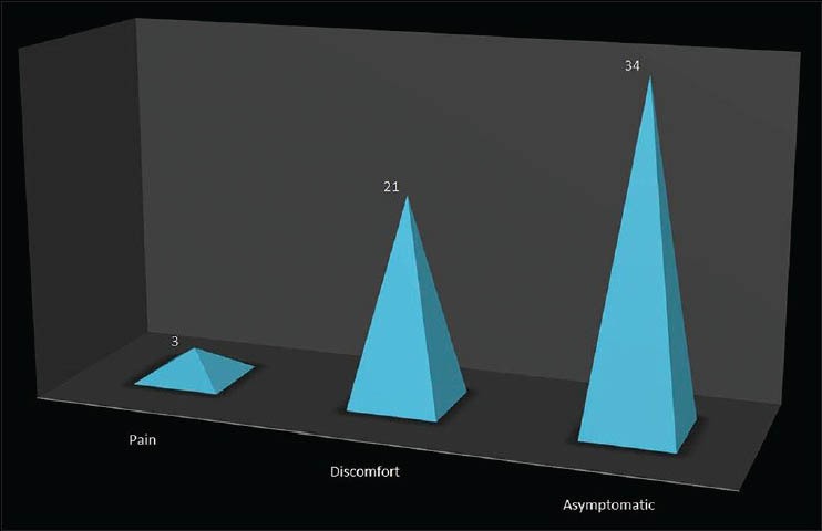
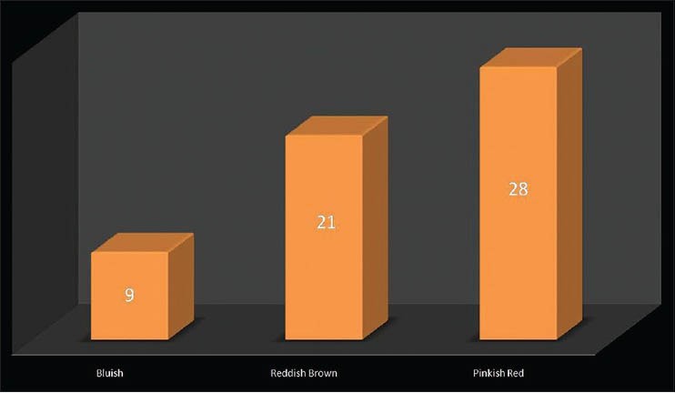
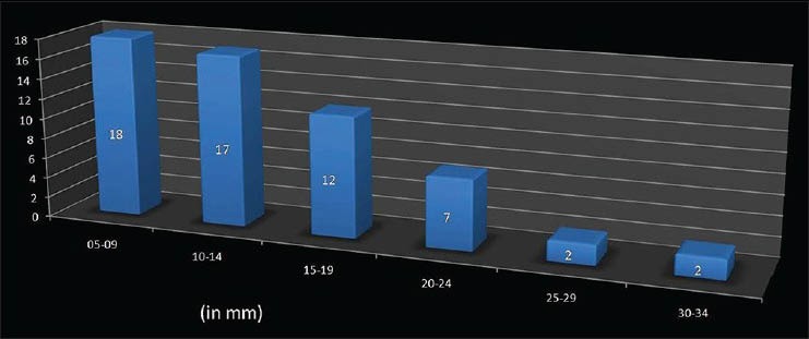
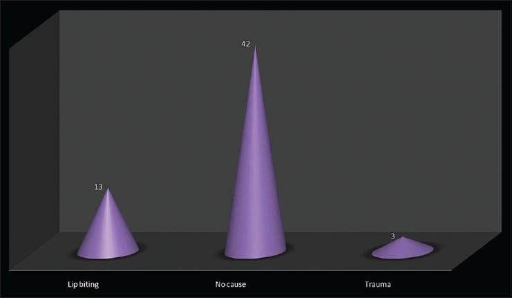
Results: Oral mucoceles were highly prevalent in the age group of 15-24 years, were seen in 51.72% of males and 48.28% of females, with a ratio of 1.07:1. The extravasation type (84.48%) was more common than the retention type (15.52%). The most common affected site was lower lip (36.20%) followed by ventral surface of the tongue (25.86%). The lowest frequency was observed in floor of mouth, upper lip and palate. The maximum numbers of mucoceles were asymptomatic (58.62%), and the color of the overlying mucosa had color of adjacent normal mucosa (48.28%). It was also observed that most of the mucoceles had diameter ranging from 5 to 14 mm. The causative factors of the lesion were lip biting (22.41%), trauma (5.18%) and numerous lesions (72.41%).
Conclusion: Oral Mucoceles are frequently seen in an oral medicine service, mainly affecting young people and lower lip, measuring around 5 to 14 mm and the extravasation type being the most common.
Keywords: Blandin and Nuhn gland; extravasation; gland of weber; oral mucocele; pseudocyst; ranula; retention; von Ebner gland.
Conflict of interest statement
Figures
References
-
- Kheur S, Desai R, Kelkar C. Mucocele of the anterior lingual salivary glands (Glands of Blandin and Nuhn) Indian J Dent Adv. 2010;2:153–5.
-
- Jani DR, Chawda J, Sundaragiri SK, Parmar G. Mucocele--A study of 36 cases. Indian J Dent Res. 2010;21:337–40. - PubMed
-
- Rashid A, Anwar N, Azizah A, Narayan K. Cases of mucocele treated in the Dental Department of Penang Hospital. Arch Orofac Sci. 2008;3:7–10.
-
- Shamim T. Oral mucocele (mucous extravasation cyst) J Ayub Med Coll Abbottabad. 2009;21:169. - PubMed
LinkOut - more resources
Full Text Sources
Other Literature Sources
