

Hydrocephalus following bilateral dumbbell-shaped c2 spinal neurofibromas resection and postoperative cervical pseudomeningocele in a patient with neurofibromatosis type 1: a case report
- PMID: 25364327
- PMCID: PMC4212698
- DOI: 10.1055/s-0034-1387805
Hydrocephalus following bilateral dumbbell-shaped c2 spinal neurofibromas resection and postoperative cervical pseudomeningocele in a patient with neurofibromatosis type 1: a case report
Abstract
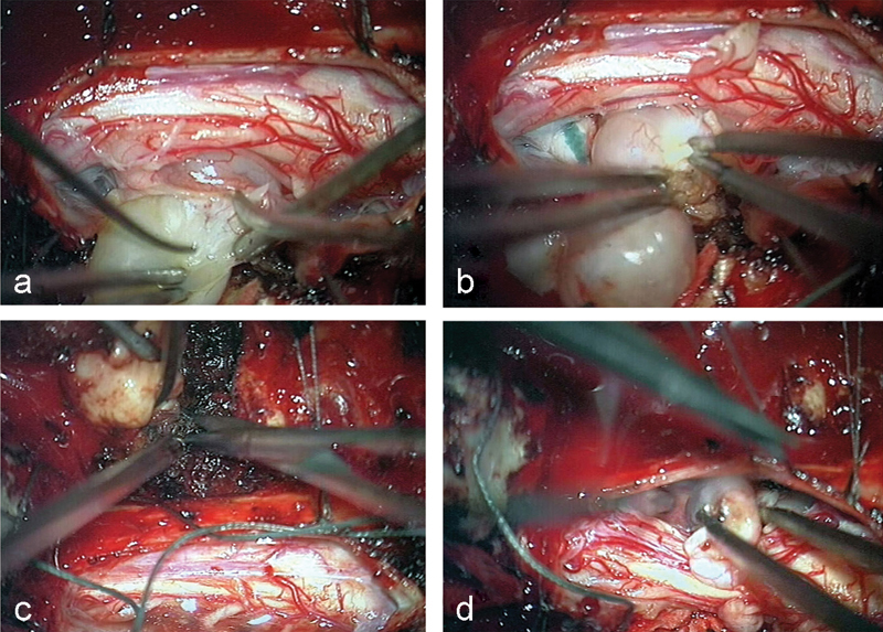
Study Design Case report. Objective To present a rare case of hydrocephalus following bilateral dumbbell-shaped C2 spinal neurofibromas resection and postoperative cervical pseudomeningocele in a patient with neurofibromatosis type 1 (NF1). Methods The patient's clinical course is retrospectively reviewed. A 37-year-old man affected by NF1 referred to our department for progressive weakness of both lower extremities and gait disturbance. Radiological imaging showed bilateral dumbbell-shaped C2 spinal neurofibromas. After its resection, at the 1-month follow-up evaluation, the patient reported headache and nausea. A CT brain scan showed a postoperative cervical pseudomeningocele and an increase in the ventricular sizes, resulting in hydrocephalus. Results A ventriculoperitoneal shunting was performed using a programmable valve opening pressure set to 120 mmH20. After surgery, the patient's neurological status markedly improved. Conclusion Hydrocephalus must be considered a possible complication of cervical spine tumor resection.
Keywords: cervical spine; dumbbell-shaped neurofibroma; hydrocephalus; neurofibromatosis type 1; postoperative cervical pseudomeningocele; spinal cord tumor.
Conflict of interest statement
Figures


Similar articles
-
Commentary on: "Hydrocephalus Following Bilateral Dumbbell-Shaped C2 Spinal Neurofibromas Resection and Postoperative Cervical Pseudomeningocele in a Patient with Neurofibromatosis Type 1: A Case Report".Evid Based Spine Care J. 2014 Oct;5(2):139-40. doi: 10.1055/s-0034-1387802. Evid Based Spine Care J. 2014. PMID: 25278888 Free PMC article. No abstract available.
-
Cervical pseudomeningocele due to occult hydrocephalus.Spine (Phila Pa 1976). 2008 May 20;33(12):E394-6. doi: 10.1097/BRS.0b013e31817343f3. Spine (Phila Pa 1976). 2008. PMID: 18496335 Review.
-
C2 neurofibromas in neurofibromatosis type 1: genetic and imaging characteristics.J Neurosurg Spine. 2018 Oct 19;30(1):126-132. doi: 10.3171/2018.7.SPINE171340. Print 2019 Jan 1. J Neurosurg Spine. 2018. PMID: 30485203
-
Subdural fluid collection and hydrocephalus following cervical schwannoma resection: hydrocephalus resolution after spinal pseudomeningocele repair: case report.J Neurosurg Spine. 2016 Dec;25(6):762-765. doi: 10.3171/2016.5.SPINE16153. Epub 2016 Jul 8. J Neurosurg Spine. 2016. PMID: 27391399
-
Neurofibroma and Meningioma within a Single Dumbbell-Shaped Tumor at the Same Cervical Level without Neurofibromatosis: A Case Report and Literature Review.World Neurosurg. 2019 Oct;130:1-6. doi: 10.1016/j.wneu.2019.06.142. Epub 2019 Jun 27. World Neurosurg. 2019. PMID: 31254713 Review.
Cited by
-
Complications of Posterior Fusion for Atlantoaxial Instability in Children With Down Syndrome.Neurospine. 2021 Dec;18(4):778-785. doi: 10.14245/ns.2142720.360. Epub 2021 Dec 31. Neurospine. 2021. PMID: 35000332 Free PMC article.
References
-
- Conference N IHCD; National Institutes of Health Consensus Development Conference. Neurofibromatosis. Conference statement Arch Neurol 1988455575–578. - PubMed
-
- Bartolomei J C Crockard H A Bilateral posterolateral approach to mirror-image C-2 neurofibromas. Report of four cases J Neurosurg 200194(2, Suppl)292–298. - PubMed
-
- Taleb F S, Guha A, Arnold P M, Fehlings M G, Massicotte E M. Surgical management of cervical spine manifestations of neurofibromatosis Type 1: long-term clinical and radiological follow-up in 22 cases. J Neurosurg Spine. 2011;14(3):356–366. - PubMed
-
- Junming M, Cheng Y, Dong C. et al.Giant cell tumor of the cervical spine: a series of 22 cases and outcomes. Spine (Phila Pa 1976) 2008;33(3):280–288. - PubMed
Publication types
LinkOut - more resources
Full Text Sources
Other Literature Sources
Research Materials
Miscellaneous
