Review
. 2014 Aug 28:2:45.
doi: 10.3389/fcell.2014.00045. eCollection 2014.

Cellular prion protein and NMDA receptor modulation: protecting against excitotoxicity

Affiliations
Review

Cellular prion protein and NMDA receptor modulation: protecting against excitotoxicity

Stefanie A G Black et al. Front Cell Dev Biol. .

Abstract

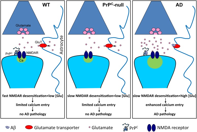
Although it is well established that misfolding of the cellular prion protein (PrP(C)) into the β-sheet-rich, aggregated scrapie conformation (PrP(Sc)) causes a variety of transmissible spongiform encephalopathies (TSEs), the physiological roles of PrP(C) are still incompletely understood. There is accumulating evidence describing the roles of PrP(C) in neurodegeneration and neuroinflammation. Recently, we identified a functional regulation of NMDA receptors by PrP(C) that involves formation of a physical protein complex between these proteins. Excessive NMDA receptor activity during conditions such as ischemia mediates enhanced Ca(2+) entry into cells and contributes to excitotoxic neuronal death. In addition, NMDA receptors and/or PrP(C) play critical roles in neuroinflammation and glial cell toxicity. Inhibition of NMDA receptor activity protects against PrP(Sc)-induced neuronal death. Moreover, in mice lacking PrP(C), infarct size is increased after focal cerebral ischemia, and absence of PrP(C) increases susceptibility of neurons to NMDA receptor-dependent death. Recently, PrP(C) was found to be a receptor for oligomeric beta-amyloid (Aβ) peptides, suggesting a role for PrP(C) in Alzheimer's disease (AD). Our recent findings suggest that Aβ peptides enhance NMDA receptor current by perturbing the normal copper- and PrP(C)-dependent regulation of these receptors. Here, we review evidence highlighting a role for PrP(C) in preventing NMDA receptor-mediated excitotoxicity and inflammation. There is a need for more detailed molecular characterization of PrP(C)-mediated regulation of NMDA receptors, such as determining which NMDA receptor subunits mediate pathogenic effects upon loss of PrP(C)-mediated regulation and identifying PrP(C) binding site(s) on the receptor. This knowledge will allow development of novel therapeutic interventions for not only TSEs, but also for AD and other neurodegenerative disorders involving dysfunction of PrP(C).

Keywords: Alzheimer's disease; NMDA receptor; beta-amyloid; cellular prion protein; excitotoxicity; ischemia; neuroinflammation.


Figures

Figure 1
Figure 1
Schematic representation of PrPC. Numbering of amino acid residues corresponds to the mouse PrPC sequence. Shown are the locations of histidine (H) residues that bind copper ions (residues 60, 68, 76, 84, 95, 110), the amino (N)-terminal signal peptide (SP; residues 1–22), β-amyloid oligomer (Aβ) binding regions (residues 23–27, 95–110), the octapeptide repeat region (OR; residues 51–90), the central hydrophobic domain (HD; residues 111–130), α-helical regions (α; residues 143–152, 171–191, 199–221), β-sheet regions (β; residues 127–129, 166–168), sites of asparagine (N)-linked glycosylation (residues 180, 196), and the carboxyl (C)-terminal glycosylphosphatidylinositol (GPI) signal peptide (GSP; residues 231–254).
Figure 2
Figure 2
Slowed NMDA receptor desensitization is pathological only in conditions of prolonged excess of glutamate. Under normal conditions (WT), glutamate homeostasis is unperturbed and prevents glutamate accumulation, and NMDA receptors (NMDAR) undergo fast desensitization, thus limiting calcium entry. In the absence of PrPC (PrPC-null), NMDA receptor desensitization is slowed but glutamate homeostasis remains unperturbed, thus limited calcium entry occurs. However, in Alzheimer's disease (AD), Aβ oligomers bind to PrPC and cause slowed NMDA receptor desensitization. This, in combination with Aβ-induced elevation of glutamate levels, for example by inhibiting glutamate re-uptake by astrocytic glutamate transporters, leads to enhanced calcium entry and pathology.

References

    1. Adle-Biassette H., Verney C., Peoc'h K., Dauge M. C., Razavi F., Choudat L., et al. (2006). Immunohistochemical expression of prion protein (PrPC) in the human forebrain during development. J. Neuropathol. Exp. Neurol. 65, 698–706. 10.1097/01.jnen.0000228137.10531.72 - DOI - PubMed
    1. Affaticati P., Mignen O., Jambou F., Potier M., Klingel-Schmitt I., Degrouard J., et al. (2011). Sustained calcium signalling and caspase-3 activation involve NMDA receptors in thymocytes in contact with dendritic cells. Cell Death Differ. 18, 99–108. 10.1038/cdd.2010.79 - DOI - PMC - PubMed
    1. Aguzzi A., Baumann F., Bremer J. (2008). The prion's elusive reason for being. Annu. Rev. Neurosci. 31, 439–477. 10.1146/annurev.neuro.31.060407.125620 - DOI - PubMed
    1. Akiyama H., Barger S., Barnum S., Bradt B., Bauer J., Cole G., et al. (2000). Inflammation and Alzheimer's disease. Neurobiol. Aging 21, 383–421. 10.1016/S0197-4580(00)00124-X - DOI - PMC - PubMed
    1. Altmeppen H. C., Prox J., Puig B., Dohler F., Falker C., Krasemann S., et al. (2013). Roles of endoproteolytic alpha-cleavage and shedding of the prion protein in neurodegeneration. FEBS J. 280, 4338–4347. 10.1111/febs.12196 - DOI - PubMed