. 2014 Nov 3;9(11):e111767.
doi: 10.1371/journal.pone.0111767. eCollection 2014.

Fibroblast growth factor 21 improves insulin sensitivity and synergizes with insulin in human adipose stem cell-derived (hASC) adipocytes

Affiliations

Fibroblast growth factor 21 improves insulin sensitivity and synergizes with insulin in human adipose stem cell-derived (hASC) adipocytes

Darwin V Lee et al. PLoS One. .

Abstract

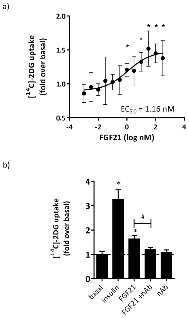
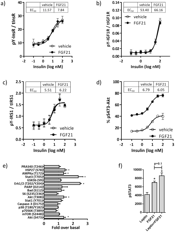
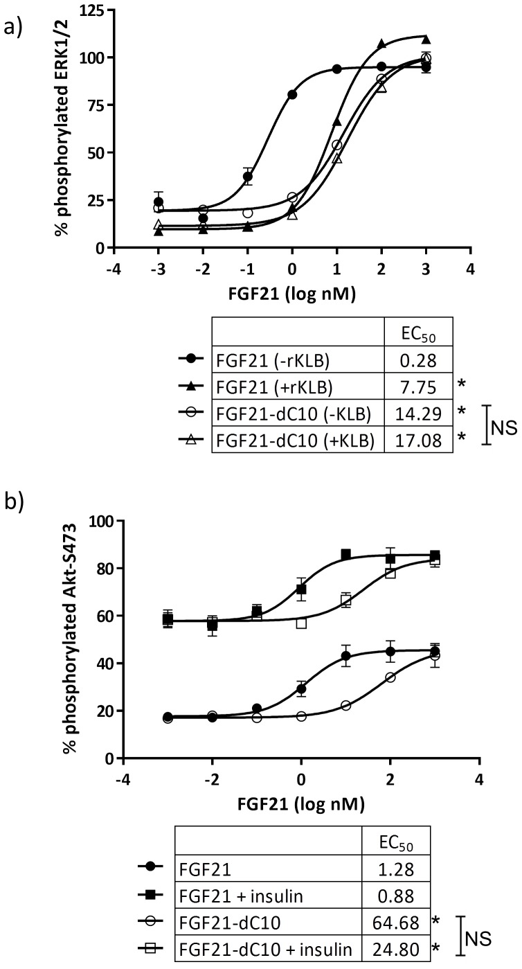
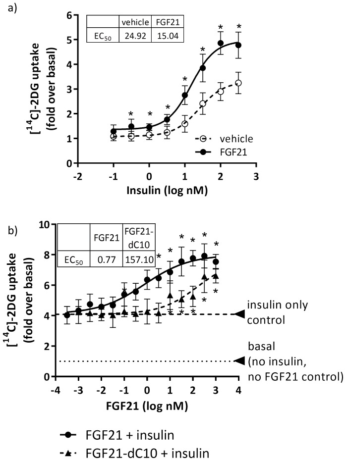
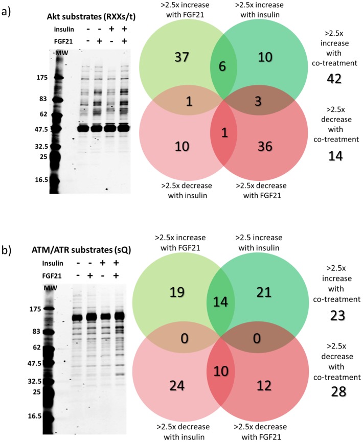
Fibroblast growth factor 21 (FGF21) has evolved as a major metabolic regulator, the pharmacological administration of which causes weight loss, insulin sensitivity and glucose control in rodents and humans. To understand the molecular mechanisms by which FGF21 exerts its metabolic effects, we developed a human in vitro model of adipocytes to examine crosstalk between FGF21 and insulin signaling. Human adipose stem cell-derived (hASC) adipocytes were acutely treated with FGF21 alone, insulin alone, or in combination. Insulin signaling under these conditions was assessed by measuring tyrosine phosphorylation of insulin receptor (InsR), insulin receptor substrate-1 (IRS-1), and serine 473 phosphorylation of Akt, followed by a functional assay using 14C-2-deoxyglucose [14C]-2DG to measure glucose uptake in these cells. FGF21 alone caused a modest increase of glucose uptake, but treatment with FGF21 in combination with insulin had a synergistic effect on glucose uptake in these cells. The presence of FGF21 also effectively lowered the insulin concentration required to achieve the same level of glucose uptake compared to the absence of FGF21 by 10-fold. This acute effect of FGF21 on insulin signaling was not due to IR, IGF-1R, or IRS-1 activation. Moreover, we observed a substantial increase in basal S473-Akt phosphorylation by FGF21 alone, in contrast to the minimal shift in basal glucose uptake. Taken together, our data demonstrate that acute co-treatment of hASC-adipocytes with FGF21 and insulin can result in a synergistic improvement in glucose uptake. These effects were shown to occur at or downstream of Akt, or separate from the canonical insulin signaling pathway.


Conflict of interest statement

Competing Interests: The study was paid for by commercial funder Pfizer, CVMED. The funder provided support in the form of salaries for authors [D.V.L., D.L., Q.Y., Y.Z., B.G., R.C., M.B.B., S.T.]. This does not alter the authors' adherence to PLOS ONE policies on sharing data and materials.

Figures

Figure 1
Figure 1. Expression of FGFR1 and KLB are consistent in hASCs and human adipose tissue.
FGFR1 and KLB mRNA in a) hASC-adipocytes, b) human subcutaneous (SC) and visceral (Vis) adipose tissue. c) Ratio of KLB:FGFR1 in human SC and VIS adipose tissue. n = 7 lean normal, n = 6 obese non T2D and n = 10 obese T2D.
Figure 2
Figure 2. FGF21 modestly induced glucose uptake, which was inhibited by anti-FGF21 neutralizing antibody.
hASC-adipocytes were treated with FGF21 for 30 minutes prior to the addition of [14C]-2DG for 30 min. a) Dose dependent effects of FGF21 on glucose uptake. N = 8 wells+S.D.; EC50 of 1.16 nM and max induction of 65% above basal signal. b) Effects of a FGF21 neutralizing antibody on the FGF21-dependent effects on glucose uptake. N = 6 wells+S.D.; induction with 32 nM FGF21 at 63% above basal, which is significantly inhibited by co-treatment with 100 ug/ml anti-FGF21 neutralizing antibody (nAb) (# p<0.05). * p<0.05 vs basal.
Figure 3
Figure 3. Activation of the insulin signaling pathway by FGF21 occurs downstream of IRS-1.
hASC-adipocytes were treated with insulin, without (veh, open-circles) or with 100 nM FGF21 (closed-circles) for 5 min, then assayed for a) insulin receptor tyrosine phosphorylation, b) IGF1R tyrosine phosphorylation, c) IRS1 tyrosine phosphorylation, and d) Akt serine-473 phosphorylation. The level of each phosphoprotein was normalized to their respective total protein content. Each value represents mean±S.D. from 3 replicate wells. d) Effects of insulin with or without FGF21 cotreatment on signaling. Insulin EC50 is displayed. e) PathScan analyses of intracellular proteins in absence and presence of 100 nM FGF21 treated-hASC-adipocytes for 10 min. *p<0.05 vs basal. f) Phosphorylation of STAT3 upon treatment with leptin alone, FGF21 alone, or in combination.
Figure 4
Figure 4. The C-terminal beta-klotho binding domain of FGF21 influenced the phosphorylation of S473-Akt by insulin and FGF21 co-treatment.
hASC-adipocytes were treated with various concentrations of FGF21 (closed-shapes) or FGF21 dC10 (open-shapes), without (circles) or with 100nM insulin (squares) for 5 min, then assayed for S473-Akt phosphorylation normalized to total Akt. Data are mean±SD for 3 replicate. * p<0.05 FGF21 vs FGF21 dC10. * Significant at p<0.05.
Figure 5
Figure 5. Synergistic induction of glucose uptake by FGF21 and insulin co-treatment was influenced by the C-terminal beta-klotho binding domain of FGF21.
a) hASC-adipocytes were treated with insulin, with or without 100 nM FGF21, for 30 minutes, then spiked with [14C]-2DG for 30 min. * p<0.05 vs vehicle. b) hASC-adipocytes were treated with FGF21 or FGF21 dC10, with or without 100 nM insulin, for 30 minutes, then spiked with [14C]-2DG for 30 min. * p<0.05 vs 100 nM insulin only. All data are mean±SD (n = 8).
Figure 6
Figure 6. FGF21 and insulin synergistically induced phosphorylation of Akt substrates and ATM/ATR substrates.
hASC-adipocytes were treated with FGF21 (10 nM) either alone or in combination with insulin (10 nM) for 30 min prior to harvest. hASC-adipocytes. a) Levels of phosphorylation of Akt substrates were assessed by western blot using a phospho-specific antibody recognizing proteins with the motif RXX(s/t). b) Levels of phosphorylation of ATM/ATR substrates were determined using a phospho-specific antibody recognizing proteins with the motif sQ.

References

    1. Kharitonenkov A, Shiyanova TL, Koester A, Ford AM, Micanovic R, et al. (2005) FGF-21 as a novel metabolic regulator. J Clin Invest 115: 1627–1635. - PMC - PubMed
    1. Fon Tacer K, Bookout AL, Ding X, Kurosu H, John GB, et al. (2010) Research resource: Comprehensive expression atlas of the fibroblast growth factor system in adult mouse. Mol Endocrinol 24: 2050–2064. - PMC - PubMed
    1. Kliewer SA, Mangelsdorf DJ (2010) Fibroblast growth factor 21: from pharmacology to physiology. Am J Clin Nutr 91: 254S–257S. - PMC - PubMed
    1. Xu J, Lloyd DJ, Hale C, Stanislaus S, Chen M, et al. (2009) Fibroblast growth factor 21 reverses hepatic steatosis, increases energy expenditure, and improves insulin sensitivity in diet-induced obese mice. Diabetes 58: 250–259. - PMC - PubMed
    1. Kharitonenkov A, Beals JM, Micanovic R, Strifler BA, Rathnachalam R, et al. (2013) Rational design of a fibroblast growth factor 21-based clinical candidate, LY2405319. PLoS One 8: e58575. - PMC - PubMed

Publication types