. 2014 Nov 3;9(11):e111591.
doi: 10.1371/journal.pone.0111591. eCollection 2014.

Bioartificial heart: a human-sized porcine model--the way ahead

Affiliations

Bioartificial heart: a human-sized porcine model--the way ahead

Alexander Weymann et al. PLoS One. .

Abstract

Background: A bioartificial heart is a theoretical alternative to transplantation or mechanical left ventricular support. Native hearts decellularized with preserved architecture and vasculature may provide an acellular tissue platform for organ regeneration. We sought to develop a tissue-engineered whole-heart neoscaffold in human-sized porcine hearts.

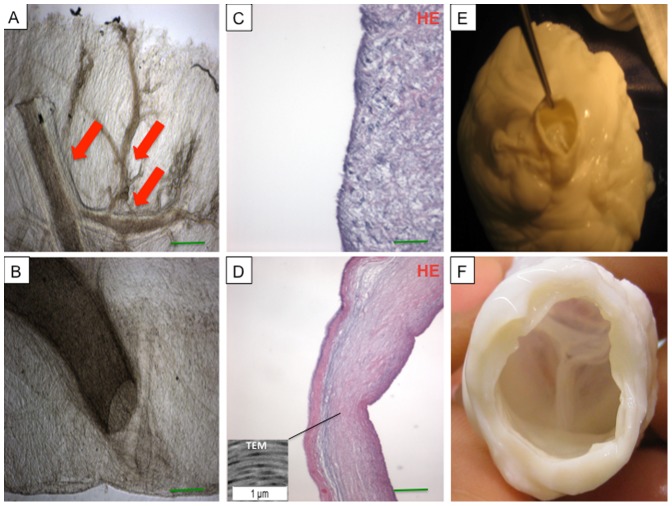
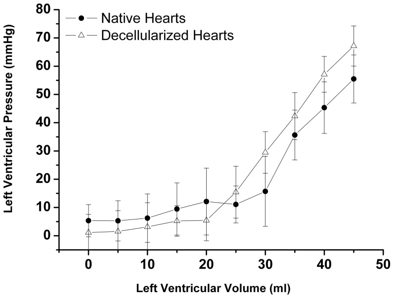
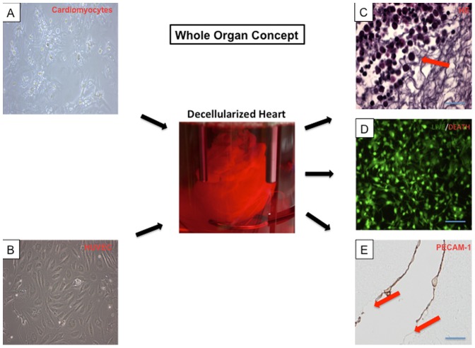
Methods: We decellularized porcine hearts (n = 10) by coronary perfusion with ionic detergents in a modified Langendorff circuit. We confirmed decellularization by histology, transmission electron microscopy and fluorescence microscopy, quantified residual DNA by spectrophotometry, and evaluated biomechanical stability with ex-vivo left-ventricular pressure/volume studies, all compared to controls. We then mounted the decellularized porcine hearts in a bioreactor and reseeded them with murine neonatal cardiac cells and human umbilical cord derived endothelial cells (HUVEC) under simulated physiological conditions.

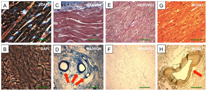
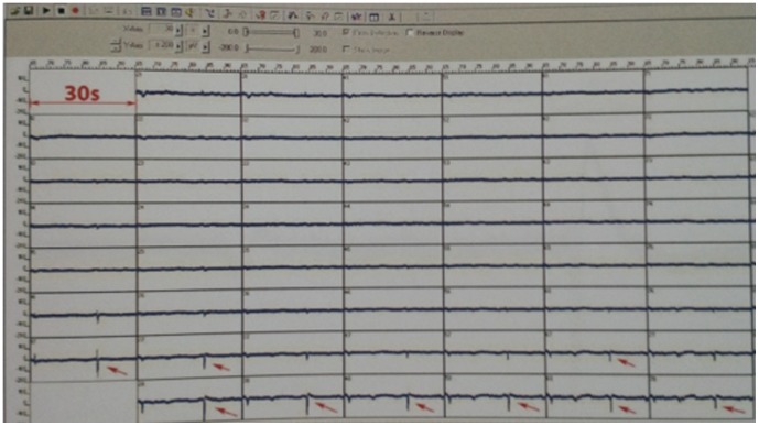
Results: Decellularized hearts lacked intracellular components but retained specific collagen fibers, proteoglycan, elastin and mechanical integrity; quantitative DNA analysis demonstrated a significant reduction of DNA compared to controls (82.6±3.2 ng DNA/mg tissue vs. 473.2±13.4 ng DNA/mg tissue, p<0.05). Recellularized porcine whole-heart neoscaffolds demonstrated re-endothelialization of coronary vasculature and measurable intrinsic myocardial electrical activity at 10 days, with perfused organ culture maintained for up to 3 weeks.

Conclusions: Human-sized decellularized porcine hearts provide a promising tissue-engineering platform that may lead to future clinical strategies in the treatment of heart failure.


Conflict of interest statement

Competing Interests: The authors have declared that no competing interests exist.

Figures

Figure 1
Figure 1. Whole-heart bioreactor: BIOSTAT B-DCU II and BioPAT DCU control tower (Sartorius Stedim Biotech GmbH, Germany).
Figure 2
Figure 2. Representative images of a porcine heart before (A) and after (B) decellularization with sodium dodecyl sulfate (SDS).
All structures including the coronary vasculature (B, red arrow) are preserved. Hematoxylin and eosin (HE) staining of ventricular tissue before (C) and after perfusion decellularization (D) showing no remnant nuclear structures after treatment with SDS, with maintained extracellular matrix and coronary vessels (D, black arrow). Scale bars, 200 µm.
Figure 3
Figure 3. Images of native (A, C, E, G) and decellularized ventricular tissue (B, D, F, H) stained with DAPI, Masson's Trichrome Stain (Masson), Herovici's Stain (Herovici) and Movat's Pentachrome Stain (Movat) revealed absence of nuclear staining after decellularization (B) and stable preservation of extracellular matrix collagen, elastic fibers including large coronary vessels (D, H, red arrow) after the decellularization procedure.
Scale bars, 200 µm.
Figure 4
Figure 4. Photomicrographs of unstained tissue samples demonstrating intact coronary vasculature (A, B) with intact third- and fourth-level vessels (A, red arrows).
The extracellular matrix composition of the aortic wall (C) and aortic valve leaflet (D) was preserved after decellularization and showed no remnant nuclear material as demonstrated by hematoxylin and eosin (HE) staining (C, D) and TEM analysis (box in D). Also the aortic valve remained competent after decellularization (E, F).
Figure 5
Figure 5. Results of biomechanical measurements.
Left ventricular peak pressure vs. volume. Decellularized hearts showed similar mechanical stability as native hearts with no significant differences in biomechanical behavior. All values are expressed as mean ± SEM.
Figure 6
Figure 6. Whole organ concept.
Cardiomyocytes (A, 60× magnification) and human umbilical vein endothelial cells (HUVEC) (B, 60× magnification) were reseeded in the decellularized porcine heart (center). Histological analysis demonstrated neonatal cardiomyocytes around the injection sites in the left ventricular wall (C, red arrow). Live/death assay demonstrated viability of the reseeded neonatal cardiomyocytes in the cultured bioartificial hearts (D). A de novo layer of endothelial cells was generated, indicated by positive PECAM-1 staining (E, cross section left coronary artery). However, the new surface cell layer of endothelial cells was partially interrupted (E, red arrows). Scale bars, 200 µm.
Figure 7
Figure 7. Demonstration of multi-electrode array electric voltage undulations of up to 200 mV in a time scale from ca. 500–1000 ms (red arrows) as a measure of myocardial electrical activity.

Similar articles

Cited by

References

    1. Kobashigawa JA, Patel JK (2006) Immunosuppression for heart transplantation: Where are we now? Nat Clin Pract Cardiovasc Med 3: 203–212. - PubMed
    1. Tonsho M, Michel S, Ahmed Z, Alessandrini A, Madsen JC (2014) Heart transplantation: Challenges facing the field. Cold Spring Harb Perspect Med 4 - PMC - PubMed
    1. Badylak SF, Freytes DO, Gilbert TW (2009) Extracellular matrix as a biological scaffold material: Structure and function. Acta biomaterialia 5: 1–13. - PubMed
    1. Weymann A, Dohmen PM, Grubitzsch H, Dushe S, Holinski S, et al. (2010) Clinical experience with expanded use of the Ross procedure: A paradigm shift? J Heart Valve Dis 19: 279–285. - PubMed
    1. Weymann A, Radovits T, Schmack B, Li S, Korkmaz S, et al. (2014) In vitro generation of atrioventricular heart valve neoscaffolds. Artif Organs 38: 118–128. - PubMed

Publication types

LinkOut - more resources