. 2015 Feb;29(2):385-93.
doi: 10.1096/fj.14-252171. Epub 2014 Nov 3.

Histone methyltransferase Dot1L plays a role in postembryonic development in Xenopus tropicalis

Affiliations

Histone methyltransferase Dot1L plays a role in postembryonic development in Xenopus tropicalis

Luan Wen et al. FASEB J. 2015 Feb.

Abstract

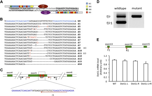
Histone methylations have been implicated to play important roles in diverse cellular processes. Of particular interest is the methylation of histone H3K79, which is catalyzed by an evolutionarily conserved methyltransferase, disruptor of telomeric silencing (Dot1)-like (Dot1L). To investigate the role of Dot1L during vertebrate development, we have generated a Dot1L-specific transcription activator-like effector nuclease (TALEN) nuclease to knockdown endogenous Dot1L in Xenopus tropicalis, a diploid species highly related to the well-known developmental model Xenopus laevis, a pseudotetraploid amphibian. We show that the TALEN was extremely efficient in mutating Dot1L when expressed in fertilized eggs, creating essentially Dot1L knockout embryos with little H3K79 methylation. Importantly, we observed that Dot1L knockdown had no apparent effect on embryogenesis because normally feeding tadpoles were formed, consistent with the lack of maternal Dot1L expression. On the other hand, Dot1L knockdown severely retarded the growth of the tadpoles and led to tadpole lethality prior to metamorphosis. These findings suggest that Dot1L and H3K79 methylation play an important role for tadpole growth and development prior to metamorphosis into a frog. Our findings further reveal interesting similarities and differences between Xenopus and mouse development and suggest the existence of 2 separate phases of vertebrate development with distinct requirements for epigenetic modifications.

Keywords: activation mark; epigenetics; histone modification; organogenesis.


Figures

Figure 1.
Figure 1.
Expression of Dot1L TALEN leads to mutations in Dot1L gene in X. tropicalis embryos. A) Schematic representation of Dot1L TALEN and the targeted DNA sequence in the Dot1L gene in X. tropicalis. The 4 types of RVDs recognizing nucleotide A, G, T, or C are depicted in different colors, respectively. The left arm contained the FokI-ELD nuclease and the right arm contained the FokI-KKR nuclease, which, when both arms bind to their respective binding sites, heterodimerize to form a functional nuclease to make a double-stranded break in the intervening sequence. B) Dot1L TALEN induces mutations in Dot1L gene. TALEN-injected embryos were used for PCR cloning of the target region in Dot1L gene and individual clones were sequenced. The sequences from mutant clones were aligned with wild-type sequence. Characters in red indicate nucleotide changes from wild-type sequence. Dashes represent nucleotide deletions. The TALEN-recognized DNA sequences were shown in blue. The ×3 shown on the right of 2 mutant sequences indicated 3 independent clones having the corresponding sequences. The number after Δ on the right indicated the number of nucleotides that were deleted in the mutant compared to the wild type. C) Schematic diagram of the Dot1L gene showing the TALEN target site and the primers used for mutant screening. The exons were shown as numbered, colored boxes. The TALEN-recognized sequences were shown as red boxes (top) or blue letters (below). Arrows represent primers. F and R were used for amplification of genomic DNA. Primers of f, r, and r1 were used for colony PCR screening for mutations. Mutations in the target region between the 2 TALEN-recognized sequences would reduce the PCR efficiency when f/r1 primers were used but would not affect the PCR by f/r primers. D) Representative PCR results from 1 wild-type and 1 mutant colony. The bacterial colonies were directly PCR amplified with primers f/r1/r. The wild-type colony produced 2 bands due to PCR amplification by f/r and f/r1, respectively. However, for the mutant, only the large band (i.e., from f/r amplification) was seen, suggesting the presence of a mutation(s) that affected the amplification by f/r1. E) Dot1L TALEN-injection does not alter total Dot1L mRNA level. Upper panel: schematic diagram of the Dot1L gene showing the locations of the primers used for RT-PCR analysis of Dot1L expression. The exons were shown as numbered, colored boxes. Lower panel: relative expression levels of Dot1L in 10-d-old tadpoles with various TALEN mRNA injections. Dot1L-L + R: injection of the mRNAs for Dot1L TALEN left and right arm. Dot1L-L: injection of TALEN Dot1L left arm mRNA plus the TALEN right arm mRNA for a control gene. Dot1L-R: injection of TALEN Dot1L right arm mRNA plus the TALEN left arm mRNA for a control gene. WT, uninjected wild-type animals.
Figure 2.
Figure 2.
There is no maternal Dot1L expression or H3K79 methylation but both are up-regulated during embryogenesis in Xenopus tropicalis. A) qRT-PCR analysis of Dot1L mRNA level during X. tropicalis development. Total RNA was isolated from embryos/tadpoles at indicated stages, and the Dot1L mRNA levels were normalized with those of the control gene ornithine decarboxylase. The expression at the early stages was magnified in the insert, showing the up-regulation of the Dot1L expression at stage 8, the midblastula stage when zygotic transcription begins. B) Western blot analysis of H3K79 dimethylation during X. tropicalis development. Note, like the lack of Dot1L mRNA at the early stages, no dimethylated H3K79 (H3K79me2) was detected until around stage 22, after the activation of zygotic Dot1L transcription (A).
Figure 3.
Figure 3.
Dot1L knockdown significantly suppressed H3K79 methylation during development. A, B) Dot1L knockdown had no observable effect on embryo morphology at 3 d (around stage 45) (A) or 6 d (stage 47) (B) postinjection. The first column is lateral view and the second is dorsal view. Numbers in the first column indicated the number of tadpoles for the sample group at 3 d. Dot1L-L + R, injection of the mRNAs for Dot1L TALEN left and right arm; Dot1L-L, injection of TALEN Dot1L left arm mRNA plus the TALEN right arm mRNA for a control gene; Dot1L-R, injection of TALEN Dot1L right arm mRNA plus the TALEN left arm mRNA for a control gene; WT, uninjected wild-type animals. C) Western blot analysis of H3K79 methylation and total [3H] in the tadpoles in (A) and (B). Note the reduction in H3K79 methylation only when the mRNAs for both Dot1L TALEN arms were injected.
Figure 4.
Figure 4.
Dot1L knockdown caused tadpole developmental retardation and lethality. A) Representative photos of tadpoles at 10 (around stage 47–49) or 16 (around stage 49) d postinjection. Note that compared to wild-type, the tadpoles were much smaller by 10 d when the mRNAs for both Dot1L TALEN arms were injected. At 10 d, the total number of animals in the TALEN injected L + R group (see Fig. 3 for more details) was 70 with a survival rate 73.0%, L group was 55 with a survival rate 93.1%, R group was 53 with a survival rate 96.4%, and WT group was 90 with a survival rate 95.7%. At 16 d postinjection, the total number of animals in the L + R group was 50 with a survival rate 55.3%, L group was 51 with a survival rate 91.3%, R group was 49 with a survival rate 94.4%, and WT group was 49 with a survival rate 93.8%. B) Developmental retardation caused by Dot1L TALEN knockdown. Tadpoles were classified into 4 categories as large (L), medium (M), small (S), and extremely small (ES) according to their sizes (see photos) at 20 d postinjection, around stage 50. The total numbers for TALEN L + R, L, R, and WT groups were 16 with a survival rate 17.1%, 45 with a survival rate 85.7%, 46 with a survival rate 94.4%, and 43 with a survival rate 89.6%, respectively. C) Tadpole survival rate over time for different TALEN injected groups. D) PCNA-immunofluorescence analysis revealed reduced cell proliferation when the mRNAs for both Dot1L TALEN arms were injected. Sections were made from the brain regions of tadpoles 17 d postinjection (around stage 49) and PCNA immunohistochemistry (green) was carried out to detect cell proliferation. The sections were also stained with DAPI (blue) for DNA. WT, uninjected wild-type animals.

Similar articles

Cited by

References

    1. Kouzarides T. (2007) Chromatin modifications and their function. Cell 128, 693–705 - PubMed
    1. Li B., Carey M., Workman J. L. (2007) The role of chromatin during transcription. Cell 128, 707–719 - PubMed
    1. Wang Z., Schones D. E., Zhao K. (2009) Characterization of human epigenomes. Curr. Opin. Genet. Dev. 19, 127–134 - PMC - PubMed
    1. Barth T. K., Imhof A. (2010) Fast signals and slow marks: the dynamics of histone modifications. Trends Biochem. Sci. 35, 618–626 - PubMed
    1. Nguyen A. T., Zhang Y. (2011) The diverse functions of Dot1 and H3K79 methylation. Genes Dev. 25, 1345–1358 - PMC - PubMed

Publication types

LinkOut - more resources