

Eradication of human immunodeficiency virus from brain reservoirs
- PMID: 25366659
- PMCID: PMC4418952
- DOI: 10.1007/s13365-014-0291-1
Eradication of human immunodeficiency virus from brain reservoirs
Abstract
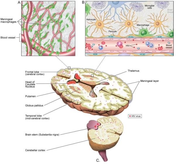
Isolated cases in which human immunodeficiency virus (HIV) infection was claimed to have been eradicated generated renewed interest in HIV reservoirs in the brain particularly since attempts to reproduce the findings using genetically engineered stem cells and immune- or myeloablation have failed. A clear understanding of the cell types in which the virus resides in the brain, the mechanism of viral persistence, restricted replication and latency, and the turnover rate of the infected cells is critical for us to develop ways to control or get rid of the virus in the brain. The brain has several unique features compared to other reservoirs. There are no resident T cells in the brain; the virus resides in macrophages and astrocytes where the viral infection is non-cytopathic. The virus evolves in the brain and since the turnover rate of these cells is low, the virus has the potential to reside in these cells for several decades and possibly for the life of the individual. This review discusses the HIV reservoirs in the brain, issues related to eradication of the virus from sanctuaries in the brain, and current challenges faced by neuroscientists in finding a cure.
Figures
References
-
- Allers K, Hutter G, Hofmann J, Loddenkemper C, Rieger K, Thiel E, et al. Evidence for the cure of HIV infection by CCR5Delta32/Delta32 stem cell transplantation. Blood. 2011;117(10):2791–9. - PubMed
-
- Hutter G, Nowak D, Mossner M, Ganepola S, Mussig A, Allers K, et al. Long-term control of HIV by CCR5 Delta32/Delta32 stem-cell transplantation. The New England journal of medicine. 2009;360(7):692–8. - PubMed
-
- Deeks SG, Wagner B, Anton PA, Mitsuyasu RT, Scadden DT, Huang C, et al. A phase II randomized study of HIV-specific T-cell gene therapy in subjects with undetectable plasma viremia on combination antiretroviral therapy. Mol Ther. 2002;5(6):788–97. - PubMed
Publication types
MeSH terms
Grants and funding
LinkOut - more resources
Full Text Sources
Other Literature Sources
Medical
