. 2014 Nov 17;211(12):2397-410.
doi: 10.1084/jem.20140625. Epub 2014 Nov 3.

Respiratory influenza virus infection induces intestinal immune injury via microbiota-mediated Th17 cell-dependent inflammation

Affiliations

Respiratory influenza virus infection induces intestinal immune injury via microbiota-mediated Th17 cell-dependent inflammation

Jian Wang et al. J Exp Med. .

Erratum in

  • J Exp Med. 2014 Dec 15;211(13):2683
  • J Exp Med. 2014 Nov 17;211(12):2396-7

Abstract

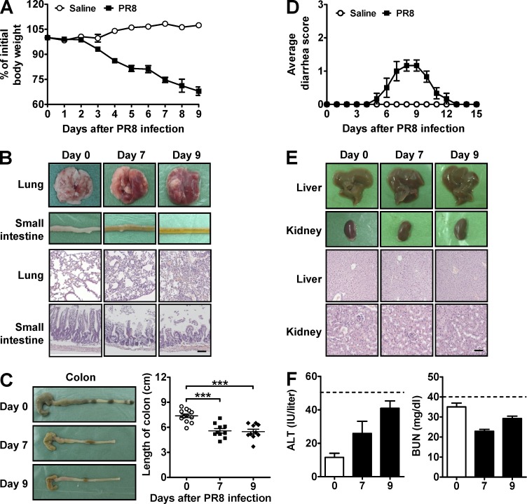
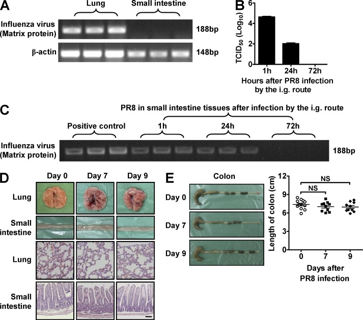
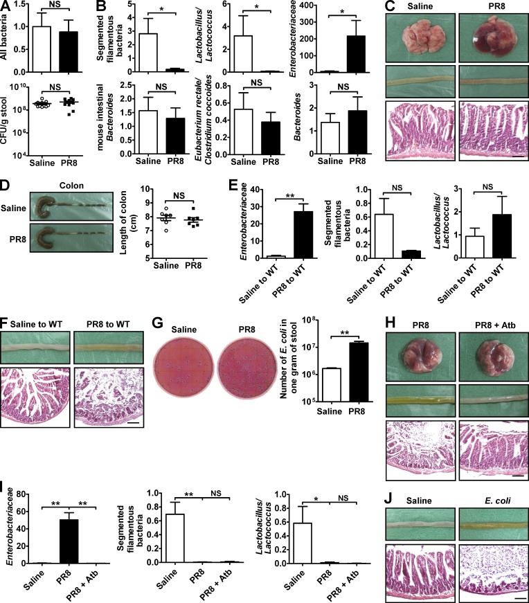
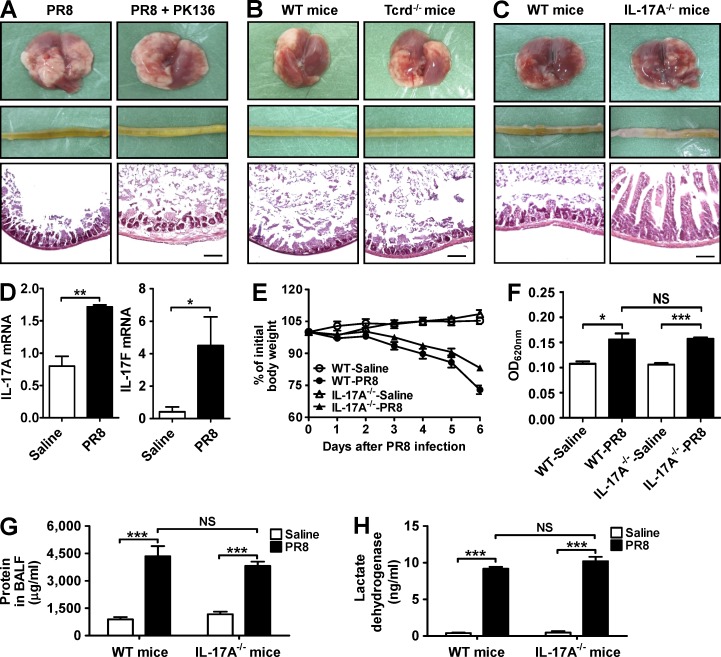
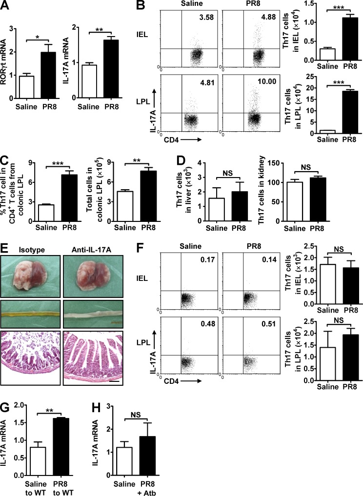
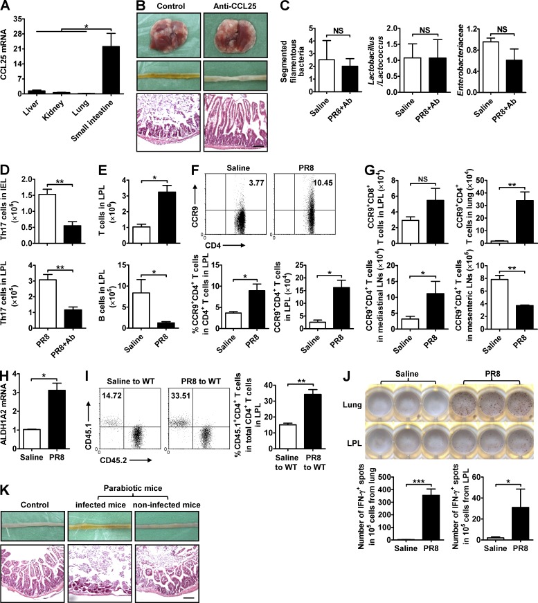
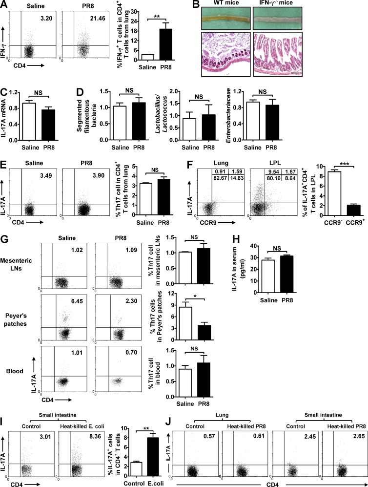
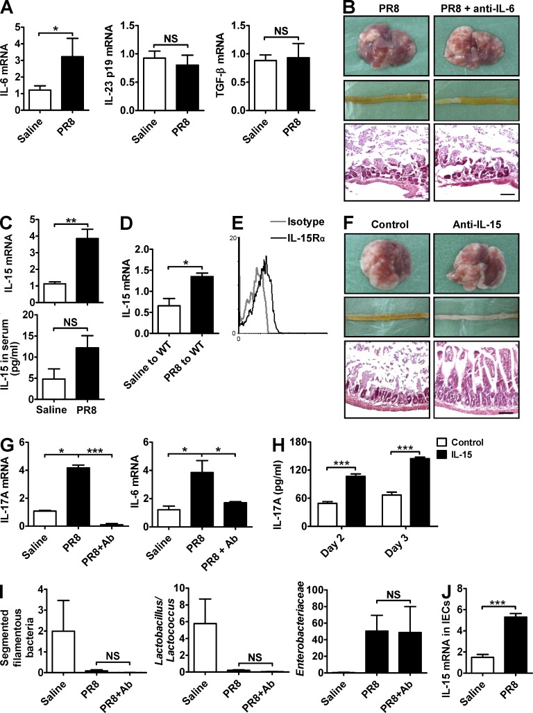
Influenza in humans is often accompanied by gastroenteritis-like symptoms such as diarrhea, but the underlying mechanism is not yet understood. We explored the occurrence of gastroenteritis-like symptoms using a mouse model of respiratory influenza infection. We found that respiratory influenza infection caused intestinal injury when lung injury occurred, which was not due to direct intestinal viral infection. Influenza infection altered the intestinal microbiota composition, which was mediated by IFN-γ produced by lung-derived CCR9(+)CD4(+) T cells recruited into the small intestine. Th17 cells markedly increased in the small intestine after PR8 infection, and neutralizing IL-17A reduced intestinal injury. Moreover, antibiotic depletion of intestinal microbiota reduced IL-17A production and attenuated influenza-caused intestinal injury. Further study showed that the alteration of intestinal microbiota significantly stimulated IL-15 production from intestinal epithelial cells, which subsequently promoted Th17 cell polarization in the small intestine in situ. Thus, our findings provide new insights into an undescribed mechanism by which respiratory influenza infection causes intestinal disease.


Figures

Figure 1.
Figure 1.
Respiratory influenza virus infection causes lung and intestinal immune injury. C57BL/6 mice were i.n. infected with saline or 0.1 HA of PR8. (A) Body weight was monitored after PR8 infection. (B) The pathology of lung and small intestine was assayed after PR8 infection. (C) The length of colon was recorded after PR8 infection. (D) The severity of the diarrhea was scored after PR8 infection (0, normal stool or absent; 1, slightly wet and soft stool; 2, wet and unformed stool with moderate perianal staining of the coat; and 3, watery stool with severe perianal staining of the coat). (E) The pathology of liver and kidney was assayed after PR8 infection. (F) Serum ALT and BUN levels were measured after PR8 infection (dashed lines represent damage threshold). All tissue sections were stained with H&E. Bars, 100 µm. Data represent three independent experiments with at least five mice/group in A, C, and D or three mice/group in B, E, and F. Data are expressed as mean ± SEM by a Student’s t test. ***, P < 0.001.
Figure 2.
Figure 2.
Influenza virus does not infect the small intestine directly. (A) C57BL/6 mice were i.n. infected with 0.1 HA of PR8. The levels of the influenza virus–derived matrix protein gene in lung and small intestine were detected by PCR. (B–E) C57BL/6 mice were i.g. infected with saline or 0.1 HA of PR8. Viral titer in intestinal contents was determined by 50% tissue culture infective dose (TCID50) assay after PR8 infection (B). The levels of the influenza virus–derived matrix protein gene in small intestine were detected by PCR after PR8 infection (C). The pathology of lung and small intestine was assayed after PR8 infection, and tissue sections were stained with H&E. Bar, 100 µm (D). The length of colon was recorded after PR8 infection (E). Data represent three independent experiments with at least three mice/group in A–E. Data are expressed as mean ± SEM by a Student’s t test. NS: not significant.
Figure 3.
Figure 3.
Antibiotic treatment reduces influenza-induced intestinal immune injury. (A) Bacteria in the small intestine were assayed by real-time PCR and selective culture in blood plate 7 d after PR8 infection. (B) Several major bacterial groups in intestinal microbiota were assayed by real-time PCR 7 d after PR8 infection. (C and D) C57BL/6 mice were subjected to a 4-wk oral treatment of combinatorial antibiotics in drinking water, followed by i.n. infection with saline or 0.1 HA of PR8. The pathology of lung and small intestine was assayed 7 d after PR8 infection (C). The length of colon was recorded 7 d after PR8 infection (D). (E and F) Transfer of intestinal microbiota from saline-treated or PR8-infected mice into healthy WT mice by the i.g. route. Major bacterial groups in the intestinal microbiota (E) and the pathology of small intestine were assayed 6 d later (F). (G) The number of E. coli in stool was detected by E. coli/Coliform Count Plates 6 d after PR8 infection. (H and I) C57BL/6 mice were subjected to a 1-wk oral treatment of streptomycin in their drinking water and then were i.n. infected with 0.1 HA of PR8. The pathology of lung and small intestine (H) and major bacterial groups in intestinal microbiota (I) were assayed 6 d after PR8 infection. (J) C57BL/6 mice were i.g. infected with saline or 5 × 10⁸ E. coli, and the pathology of small intestine was assayed 3 d later. All tissue sections were stained with H&E. Bars, 100 µm. Data represent two independent experiments with three mice/group in I and J or three independent experiments with at least three mice/group in A–H. Data are expressed as mean ± SEM by a Student’s t test. *, P < 0.05; **, P < 0.01; NS: not significant.
Figure 4.
Figure 4.
IL-17A deficiency reduces influenza-induced immune injury in small intestine but not in lung. (A) The pathology of lung and small intestine from control and PK136-treated mice was assayed 6 d after PR8 infection. (B) The pathology of lung and small intestine from WT and Tcrd−/− mice was assayed 6 d after PR8 infection. (C) The pathology of lung and small intestine from WT and IL-17A−/− mice was assayed 6 d after PR8 infection. (D) IL-17A and IL-17F expressions in the lung from WT mice were detected by real-time PCR 6 d after PR8 infection. (E) Body weight of WT and IL-17A−/− mice was monitored after PR8 infection. (F) Evans blue dye concentration in BALF from WT and IL-17A−/− mice was determined by spectrophotometer 6 d after PR8 infection. (G and H) Total protein (G) and lactate dehydrogenase (H) levels in BALF from WT and IL-17A−/− mice were determined by ELISA 6 d after PR8 infection. All tissue sections were stained with H&E. Bars, 100 µm. Data represent two independent experiments with five mice/group in E–H or three mice/group in A–D. Data are expressed as mean ± SEM by a Student’s t test. *, P < 0.05; **, P < 0.01; ***, P < 0.001; NS: not significant.
Figure 5.
Figure 5.
Increased Th17 cells occur in the small intestine during influenza virus infection. (A) RORγt and IL-17A expressions in the small intestine were detected by real-time PCR 7 d after PR8 infection. (B) The percentage and number of Th17 cells in intestinal IEL and LPL were detected 7 d after PR8 infection. (C) The percentage and number of Th17 cells in colonic LPL were detected 7 d after PR8 infection. (D) The number of Th17 cells in liver and kidney was detected 7 d after PR8 infection. (E) C57BL/6 mice were i.p. treated with a neutralizing anti–IL-17A antibody during PR8 infection. The pathology of lung and small intestine was assayed 6 d after PR8 infection, and tissue sections were stained with H&E. Bars, 100 µm. (F) The percentage and number of Th17 cells in IEL and LPL were detected 7 d after PR8 infection in antibiotic-treated mice. (G) Transfer of intestinal microbiota from saline-treated or PR8-infected mice into healthy WT mice by the i.g. route. IL-17A expression in the small intestine was detected by real-time PCR 6 d later. (H) IL-17A expression in the small intestine was detected by real-time PCR at day 6 after PR8 infection in streptomycin-treated mice. Data represent two independent experiments with three mice/group in A, D, E, G, and H or three independent experiments with three mice/group in B, C, and F. Data are expressed as mean ± SEM by a Student’s t test. *, P < 0.05; **, P < 0.01; ***, P < 0.001; NS: not significant.
Figure 6.
Figure 6.
Anti-CCL25 antibody treatment reduces influenza–induced intestinal immune injury. (A) CCL25 expression in various tissues was detected by real-time PCR 4 d after PR8 infection. (B–D) C57BL/6 mice were i.v. treated with a neutralizing anti-CCL25 antibody during PR8 infection. The pathology of lung and small intestine (B), major bacterial groups in intestinal microbiota (C), and the number of Th17 cells in IEL and LPL were assayed 7 d after PR8 infection (D). (E–G) C57BL/6 mice were i.n. infected with saline or 0.1 HA of PR8. The number of T and B cells in LPL (E), the percentage and number of CCR9+CD4+ T cells in small intestine (F), and the number of CCR9+CD8+ T cells in LPL and CCR9+CD4+ T cells in lung, mediastinal LNs, and mesenteric LNs were assayed 7 d after PR8 infection (G). (H) ALDH1A2 expression in lung was detected by real-time PCR 6 d after PR8 infection. (I) CD4+ T cells from the lungs of saline- or PR8-infected CD45.1+ mice were adoptively transferred into WT CD45.2+ mice, and the percentage of CD45.1+CD4+ T cells in total CD4+ T cells in LPL from recipient CD45.2+ mice was detected by flow cytometry 48 h later. (J) C57BL/6 mice were i.n. infected with saline or 0.1 HA of PR8. CD4+ T cells in the lung and LPL were purified 6 d later by MACS and then co-cultured with antigen-presenting cells and heat-killed PR8 in an IFN-γ ELISPOT plate. The number of positive spots was counted 20 h later. (K) Parabiotic pairs of WT mice were established first, and the left partner was i.n. infected with PR8 2 wk later. The pathology of small intestine was assayed 6 d after PR8 infection. All tissue sections were stained with H&E. Bars, 100 µm. Data represent three independent experiments with three mice/group in A–H and K or three wells/treatment in J. Data are expressed as mean ± SEM by a Student’s t test. *, P < 0.05; **, P < 0.01; ***, P < 0.001; NS: not significant.
Figure 7.
Figure 7.
Lung-derived CD4+ T cells influence microbiota and intestine injury by secreting IFN-γ. (A) IFN-γ expression in CD4+ T cells from lung was detected by flow cytometry 6 d after PR8 infection in WT mice. (B) The pathology of small intestine was assayed at day 7 after PR8 infection in WT and IFN-γ−/− mice, and tissue sections were stained with H&E. Bars, 100 µm. (C and D) IL-17A expression in the small intestine (C) and major bacterial groups in intestinal microbiota (D) were assayed at day 7 after PR8 infection in IFN-γ−/− mice. (E and F) IL-17A expression in CD4+ T cells from lung (E) and the percentages of CCR9 Th17 cells and CCR9+ Th17 cells in lung and LPL (F) were detected 6 d after PR8 infection in WT mice. (G) IL-17A expression in CD4+ T cells from mesenteric LNs, Peyer’s patches, and blood was detected by flow cytometry 6 d after PR8 infection in WT mice. (H) IL-17A level in serum was detected by ELISA 6 d after PR8 infection in WT mice. (I) LPL from WT mice at day 6 after PR8 infection was stimulated by heat-killed E. coli in vitro; 24 h later, the expression of IL-17A in CD4+ T cells was detected by flow cytometry. (J) Lung lymphocytes and LPL from WT mice at day 6 after PR8 infection were stimulated by heat-killed PR8 in vitro; 24 h later, the expression of IL-17A in CD4+ T cells was detected by flow cytometry. Data represent three independent experiments with three mice/group in A–H or three wells/treatment in I and J. Data are expressed as mean ± SEM by a Student’s t test. *, P < 0.05; **, P < 0.01; ***, P < 0.001; NS: not significant.
Figure 8.
Figure 8.
Intestinal microbiota induces Th17 cell polarization in situ via triggering IL-15 production. (A) IL-6, IL-23, and TGF-β expressions in the small intestine were detected by real-time PCR 6 d after PR8 infection. (B) The pathology of lung and small intestine from control and anti–IL-6–treated mice was assayed 6 d after PR8 infection. (C) IL-15 expression in the small intestine and serum was detected 6 d after PR8 infection. (D) Transfer of intestinal microbiota from saline-treated or PR8-infected mice into healthy WT mice by the i.g. route. IL-15 expression in the small intestine was detected 6 d later. (E) IL-15Rα expression on CD4+ T cells in LPL from WT mice was detected at day 6 after PR8 infection. (F and G) C57BL/6 mice were i.p. treated with a neutralizing anti–IL-15 antibody during PR8 infection. The pathology of lung and small intestine (F) as well as IL-17A and IL-6 expressions in the small intestine were assayed 6 d after PR8 infection (G). (H) MACS-purified CD4+ T cells from LPL were stimulated by IL-15 in vitro, and IL-17A levels in supernatant were measured at days 2 and 3 by ELISA. (I) Major bacterial groups in the intestinal microbiota from control and anti–IL-15–treated mice were assayed by real-time PCR 6 d after PR8 infection. (J) IL-15 expression in IECs was detected 6 d after PR8 infection in WT mice. All tissue sections were stained with H&E. Bars, 100 µm. Data represent three independent experiments with three mice/group in A–G, I, and J or three wells/treatment in H. Data are expressed as mean ± SEM by a Student’s t test. *, P < 0.05; **, P < 0.01; ***, P < 0.001; NS: not significant.

Comment in

  • Mucosal immunology: sick of the flu.
    Minton K. Minton K. Nat Rev Immunol. 2014 Dec;14(12):781. doi: 10.1038/nri3773. Epub 2014 Nov 14. Nat Rev Immunol. 2014. PMID: 25394944 No abstract available.
  • FLUshing in the bathroom.
    Amezcua C, Gagliani N, Flavell R. Amezcua C, et al. J Exp Med. 2014 Nov 17;211(12):2328-9. doi: 10.1084/jem.21112insight4. J Exp Med. 2014. PMID: 25403806 Free PMC article. No abstract available.

Similar articles

Cited by

References

    1. Baden L.R., Drazen J.M., Kritek P.A., Curfman G.D., Morrissey S., and Campion E.W.. 2009. H1N1 influenza A disease—information for health professionals. N. Engl. J. Med. 360:2666–2667 10.1056/NEJMe0903992 - DOI - PubMed
    1. Campbell D.J., and Butcher E.C.. 2002. Intestinal attraction: CCL25 functions in effector lymphocyte recruitment to the small intestine. J. Clin. Invest. 110:1079–1081 10.1172/JCI0216946 - DOI - PMC - PubMed
    1. Chervonsky A.2009. Innate receptors and microbes in induction of autoimmunity. Curr. Opin. Immunol. 21:641–647 10.1016/j.coi.2009.08.003 - DOI - PMC - PubMed
    1. Crowe C.R., Chen K., Pociask D.A., Alcorn J.F., Krivich C., Enelow R.I., Ross T.M., Witztum J.L., and Kolls J.K.. 2009. Critical role of IL-17RA in immunopathology of influenza infection. J. Immunol. 183:5301–5310 10.4049/jimmunol.0900995 - DOI - PMC - PubMed
    1. Das G., Augustine M.M., Das J., Bottomly K., Ray P., and Ray A.. 2003. An important regulatory role for CD4+CD8αα T cells in the intestinal epithelial layer in the prevention of inflammatory bowel disease. Proc. Natl. Acad. Sci. USA. 100:5324–5329 10.1073/pnas.0831037100 - DOI - PMC - PubMed

Publication types

MeSH terms