Review
. 2014 Nov 1;28(21):2331-47.
doi: 10.1101/gad.250647.114.

Pericellular proteolysis in cancer

Affiliations
Review

Pericellular proteolysis in cancer

Lisa Sevenich et al. Genes Dev. .

Abstract

Pericellular proteases have long been associated with cancer invasion and metastasis due to their ability to degrade extracellular matrix components. Recent studies demonstrate that proteases also modulate tumor progression and metastasis through highly regulated and complex processes involving cleavage, processing, or shedding of cell adhesion molecules, growth factors, cytokines, and kinases. In this review, we address how cancer cells, together with their surrounding microenvironment, regulate pericellular proteolysis. We dissect the multitude of mechanisms by which pericellular proteases contribute to cancer progression and discuss how this knowledge can be integrated into therapeutic opportunities.

Keywords: invasion; macrophages; metastasis; migration; proteases; tumor microenvironment.


Figures

Figure 1.
Figure 1.
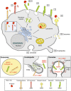
Protein trafficking of pericellular proteases. Pericellular proteases are synthesized in the endoplasmic reticulum (ER) and transported through the Golgi complex to the trans-Golgi-network (TGN). (1) Membrane-associated proteases are activated by furin in the TGN and reach the cell surface as active proteases. (2) Alternatively, membrane-associated proteases can be transported to the cell surface as inactive precursor proteins and are proteolytically activated in the pericellular space. (3) Classically secreted proteases are transported to the plasma membrane (PM) through the constitutive secretory pathway and are proteolytically activated in the pericellular space following secretion. Proteases that are typically localized to endosomes or lysosomes can be transported to the extracellular space through alternative trafficking via the secretory pathway (4) or through lysosomal exocytosis (5). Lysosomal proteases that reach the extracellular space via the secretory pathway are secreted as proenzymes and require proteolytic activation, while proteases that trafficked through the lysosome are activated within the lysosome and secreted as active enzymes. Secreted proteases can be tethered to the PM through interacting partners such as CD44, integrins, or annexin II (An II) (6) or through binding to ECM components (7). (8) Some secreted proteases are bound to specific receptors; e.g., uPA binds to uPAR. (9) Proteases can also be released through exosomes that originate from multivesicular bodies (MVBs), which leads to secretion into the extracellular space or transfer to adjacent cells. Accumulation or localized release of pericellular proteases is associated with PM microdomains such as invadopodia (actin-rich protrusions) (10) or caveolae (a subset of lipid rafts) (11), which are represented at higher magnification in the bottom panels.
Figure 2.
Figure 2.
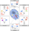
Microenvironmental regulation of pericellular proteolysis. Interactions between tumor cells and noncancerous stromal cells, including fibroblasts, inflammatory cells (e.g., macrophages, neutrophils, and dendritic cells), immune cells (e.g., B and T cells), and endothelial cells, have been reported to induce protease expression and activity within the tumor microenvironment. Paracrine signaling between tumor cells and stromal cells is orchestrated through cytokines and growth factors. Noncellular stimuli such as hypoxia or acidic extracellular pH are also known inducers of protease expression and activity.
Figure 3.
Figure 3.
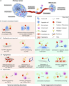
Tumor-promoting and tumor-suppressive functions of pericellular proteases in cancer. (A) Schematic overview of rate-limiting steps during primary tumor growth and metastasis that are regulated by pericellular proteases. (B) Cleavage of different substrates modulates distinct processes in primary cancer progression and metastatic dissemination. Pericellular proteases promote local invasion of tumor cells, intravasation into the circulation, and extravasation at the secondary site by cleavage of ECM components and cell adhesion molecules such as E-cadherin, JAM-B, or occludin. (C) Pericellular proteases promote tumor cell proliferation and survival by ECM processing, which liberates cytokines that are tethered to the ECM. Certain cytokines require proteolytic cleavage for their activation or bioavailability; e.g., cleavage of IGF-BP liberates IGF. Conversely, proteases can reduce tumor cell proliferation and survival by proteolytic degradation of cytokines or inactivation of cytokine receptors by ectodomain shedding. Generation of apoptotic factors (e.g., sFasL) induces apoptosis in tumor cells. (D) Pericellular proteases exert proangiogenic functions by ECM remodeling during vessel sprouting and degradation of anti-angiogenic factors such as angiostatin and tumstatin. However, more restricted proteolytic processing of ECM components can lead to the generation of these same anti-angiogenic matrikines, angiostatin and tumstatin. Thus, the balance between these activities will determine whether pericellular proteolysis is proangiogenic or anti-angiogenic. Cleavage of uPAR on the cell surface of endothelial cells also inhibits angiogenesis by limiting endothelial cell invasion. (E) Pericellular proteases regulate the activation status of different stromal cell types and orchestrate the recruitment of tumor-promoting or tumor-suppressing inflammatory cells. Proteolytic liberation of TGF-β leads to suppression of T-cell responses against tumor cells, and shedding of IL-2Rα or IFN-R leads to alterations in the activation status of T cells. Pericellular proteases are also implicated in anti-cancer immune responses, e.g., by inactivating members of the CC and CXC chemokine family, thereby blocking the recruitment of tumor-promoting inflammatory cells. Conversely, processing of chemokines may also recruit tumor-suppressive inflammatory cells.

Similar articles

Cited by

References

    1. Affara NI, Andreu P, Coussens LM. 2009. Delineating protease functions during cancer development. Methods Mol Biol 539: 1–32 - PubMed
    1. Akkari L, Gocheva V, Kester JC, Hunter KE, Quick ML, Sevenich L, Wang H-W, Peters C, Tang LH, Klimstra D, et al. . 2014. Distinct functions of macrophage- and cancer cell-derived cathepsin Z combine to promote tumor malignancy via interactions with the extracellular matrix. Genes Dev 28: 2134–2150 - PMC - PubMed
    1. Almeida PC, Nantes IL, Rizzi CC, Judice WA, Chagas JR, Juliano L, Nader HB, Tersariol IL. 1999. Cysteine proteinase activity regulation: a possible role of heparin and heparin-like glycosaminoglycans. J Biol Chem 274: 30433–30438 - PubMed
    1. Almeida PC, Nantes IL, Chagas JR, Rizzi CC, Faljoni-Alario A, Carmona E, Juliano L, Nader HB, Tersariol IL. 2001. Cathepsin B activity regulation. Heparin-like glycosaminogylcans protect human cathepsin B from alkaline pH-induced inactivation. J Biol Chem 276: 944–951 - PubMed
    1. Antalis TM, Bugge TH, Wu Q. 2011. Membrane-anchored serine proteases in health and disease. Prog Mol Biol Transl Sci 99: 1–50 - PMC - PubMed

Publication types