. 2015 Apr;473(4):1349-57.
doi: 10.1007/s11999-014-4021-z.

No regeneration of the human acetabular labrum after excision to bone

Affiliations

No regeneration of the human acetabular labrum after excision to bone

Hermes H Miozzari et al. Clin Orthop Relat Res. 2015 Apr.

Abstract

Background: Treatment options for a symptomatic, torn, irreparable, or completely ossified acetabular labrum are limited to either excision and/or reconstruction with grafts. In a previous animal model, regeneration of the acetabular labrum after excision to the bony rim has been shown. In humans, less is known about the potential of regeneration of the labrum. Recent studies seem to confirm labral regrowth, but it is still unclear if wide excision might be a surgical option in cases where repair is not possible.

Questions/purposes: The purposes of this study were (1) to determine the extent of acetabular labrum regeneration after excision to the bony rim; and (2) to determine whether this procedure results in higher hip scores.

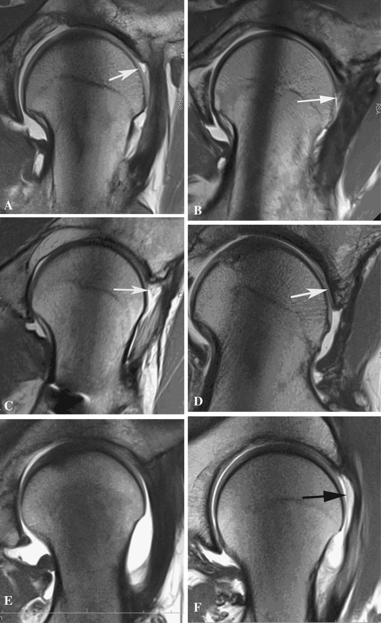
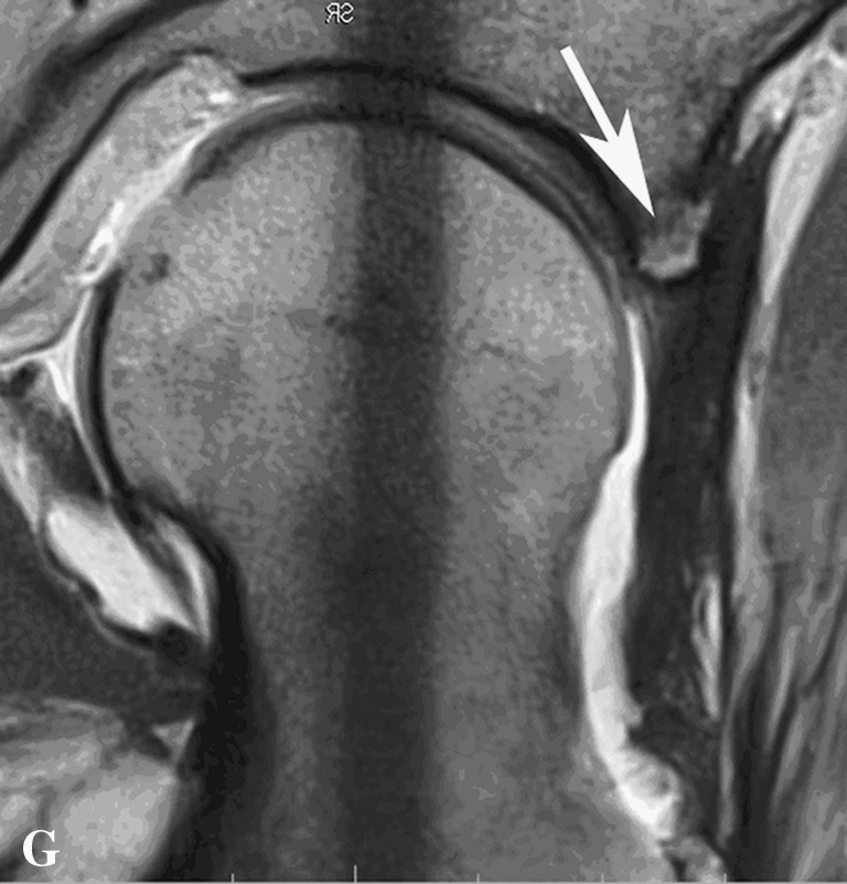
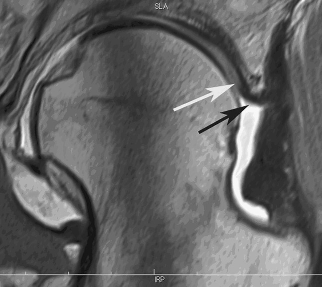
Methods: We reviewed all patients treated with surgical dislocation for symptomatic femoroacetabular impingement by a single surgeon at one institution between 2003 and 2008, of whom 14 underwent wide labral excision (of at least 60°) down to bone; we used this approach when there was an absence of reparable tissue. Of these 14, nine were available for voluntary reexamination. The mean age at surgery was 38 ± 9 SD years and the mean followup was 4 ± 1 SD years. All patients consented to a physical examination and an MRI arthrogram, which was evaluated for evidence of new tissue formation by four observers. A modified Harris hip score and the UCLA were recorded.

Results: Regrowth of a structure equivalent to normal labrum was not observed on the MRI arthrograms. Six of nine hips had segmental defects, bone formation was found in five, and the capsule was confluent with the new tissue in six. The mean Harris hip score at latest followup was 83 ± 14, and the mean UCLA score was 6 ± 2.

Conclusions: Resection of a nonreparable acetabular labrum down to a bleeding bony surface does not stimulate regrowth of tissue that appears to be capable of normal function by MR arthrography, and patients who underwent this procedure had lower hip scores at midterm than previously reported from the same institution for patients undergoing labral repair or sparse débridement. Based on these results, we believe that future studies should evaluate alternatives to reconstructing the labrum, perhaps using ligamentum teres, because resection seems neither to result in regrowth nor the restoration of consistently high hip scores.


Figures

Fig. 1A–G
Fig. 1A–G
These are illustrations of the MRA appearance of the tissue response in the area of previous resection of the acetabular labrum down to the bony rim as seen on radial views. In each case, the capsule and adjacent musculature have reconstituted after surgical dislocation and anterior arthrotomy. (A) No regrowth; a void is present between the capsule and the femoral head (white arrow) (response A). (B) Replacement with tissue (white arrow) resembling the normal labrum in density, shape, and location relative to the acetabular rim and femoral head (read as response B1). Infiltration of contrasts shows a probable tear in the new labral tissue. Major adhesions between the neck in the area of femoral osteoplasty are visible. (C) Tissue has formed in the void (white arrow) but is low-density fibrous tissue or synovium (response B2). (D) The void has been filled with bone (response B3). Where the undersurface of the bone abuts the femoral head, it is smooth and covered with soft tissue (white arrow), similar to an osteophyte. (E) The resection site is filled with dense fibrous tissue indistinguishable from the adjacent capsule and similar in density to normal labrum (response C1). (F) The void has been partially replaced with low-density tissue (black arrow) that is fused with the capsule (response C2). (G) The area of prior resection is largely replaced by bone (white arrow) that is enveloped by the capsule (response C3).
Fig. 1A–G
Fig. 1A–G
These are illustrations of the MRA appearance of the tissue response in the area of previous resection of the acetabular labrum down to the bony rim as seen on radial views. In each case, the capsule and adjacent musculature have reconstituted after surgical dislocation and anterior arthrotomy. (A) No regrowth; a void is present between the capsule and the femoral head (white arrow) (response A). (B) Replacement with tissue (white arrow) resembling the normal labrum in density, shape, and location relative to the acetabular rim and femoral head (read as response B1). Infiltration of contrasts shows a probable tear in the new labral tissue. Major adhesions between the neck in the area of femoral osteoplasty are visible. (C) Tissue has formed in the void (white arrow) but is low-density fibrous tissue or synovium (response B2). (D) The void has been filled with bone (response B3). Where the undersurface of the bone abuts the femoral head, it is smooth and covered with soft tissue (white arrow), similar to an osteophyte. (E) The resection site is filled with dense fibrous tissue indistinguishable from the adjacent capsule and similar in density to normal labrum (response C1). (F) The void has been partially replaced with low-density tissue (black arrow) that is fused with the capsule (response C2). (G) The area of prior resection is largely replaced by bone (white arrow) that is enveloped by the capsule (response C3).
Fig. 2
Fig. 2
Here the void has been partially filled with new bone (white arrow), which is covered by a layer of dense fibrous tissue (black arrow) that could appear to be normal labral edge by arthroscopy.

Comment in

References

    1. Abrams GD, Safran MR, Sadri H. Spontaneous hip labrum regrowth after initial surgical débridement. Clin Orthop Relat Res. 2013;471:2504–2508. doi: 10.1007/s11999-013-2914-x. - DOI - PMC - PubMed
    1. Altenberg AR. Acetabular labrum tears: a cause of hip pain and degenerative arthritis. South Med J. 1977;70:174–175. doi: 10.1097/00007611-197702000-00019. - DOI - PubMed
    1. Aprato A, Jayasekera N, Villar RN. The accuracy of magnetic resonance arthrography in hip arthroscopic labral revision surgery. Hip Int. 2013;23:99–103. doi: 10.5301/HIP.2012.10436. - DOI - PubMed
    1. Aprato A, Jayasekera N, Villar RN. Revision hip arthroscopic surgery: outcome at three years. Knee Surg Sports Traumatol Arthrosc. 2014;22:932–937. doi: 10.1007/s00167-013-2373-7. - DOI - PubMed
    1. Aprato A, Masse A, Faletti C, Valente A, Atzori F, Stratta M, Jayasekera N. Magnetic resonance arthrography for femoroacetabular impingement surgery: is it reliable? J Orthop Traumatol. 2013;14:201–206. doi: 10.1007/s10195-013-0227-1. - DOI - PMC - PubMed