

doi: 10.1038/cr.2014.141.
Epub 2014 Nov 4.
EGF promotes mammalian cell growth by suppressing cellular senescence
Affiliations
- PMID: 25367123
- PMCID: PMC4650583
- DOI: 10.1038/cr.2014.141
Item in Clipboard
EGF promotes mammalian cell growth by suppressing cellular senescence
Cell Res.
2015 Jan.
No abstract available
Figures
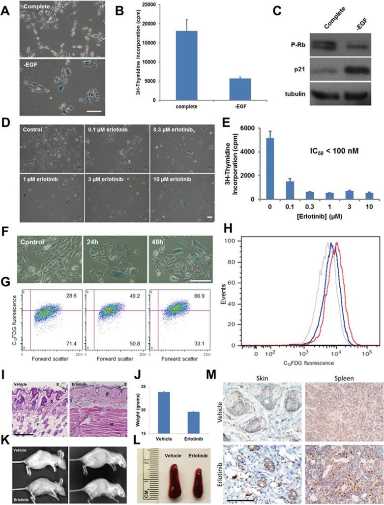
Anti-senescence function of EGF in human epithelial cells and mouse tissues. (A-C) EGF depletion results in cellular senescence of HME cells. HME cells were exposed for 1 week to complete culture medium or to medium lacking EGF. Cells cultured in the absence of EGF displayed an enlarged morphology, elevated SA-β-gal activity (A), decreased proliferation (B), and reduced Rb phosphorylation and elevated p21 expression (C), all markers that are commonly associated with cellular senescence. (D-H) EGFR pharmacological inhibition causes cellular senescence of HME cells. In D and E, HME cells were treated for three days with complete culture medium supplemented with increasing concentrations of erlotinib. Cells were then stained for SA-β-gal activity (D), or metabolically labeled with 2 μCi of [3H]-thymidine for 16 h (E). Progressively decreasing amounts of label incorporation were observed for cells cultured in the presence of erlotinib. Experiments were performed in triplicate; error bars indicate SD. In F-H, quantification of erlotinib-induced cellular senescence of HME cells using flow cytometry. HME cells were exposed to 1 μM erlotinib for 24 or 48 h and then analyzed for SA-β-gal activity using either a chromogenic assay (F) or a fluorescence-based assay (G and H, see Supplementary information, Data S1). Both assay types indicated that SA-β-gal reached maximal activity from 24 to 48 h after treatment. (A, D, F) Scale bar, 50 μm. (I-M) Mice treated with erlotinib display early aging-associated phenotypes. Skin atrophy in erlotinib-treated mice. BALB/c nude mice (five per group) were treated with either vehicle or erlotinib (50 mg/kg/day) for 4 weeks. Skin was fixed in 4% PFA, stained using H&E, and photographed at 10× magnification. Note the reduced epidermal thickness and fat content resulting from EGFR inhibition. E, epidermis; D, dermis; F, fat; M, muscle. Scale bar, 250 μm. (I). Reduced weight of mice treated with daily erlotinib. Error bars indicate SD (J). Representative photograph and X-ray of tumor-bearing BALB/c nude mice treated with vehicle or erlotinib. Note the reduced size, kyphosis, and generally aged appearance of the erlotinib-treated mouse (K). Spleen atrophy in erlotinib-treated mice (see also Supplementary information, Figure S1P-S1Q) (L). Immunohistochemical staining of skin and spleen tissue from vehicle- or erlotinib-treated mice was performed using an antibody specific for p16. Images shown were photographed at 40×. Scale bar, 100 μm (M).
Similar articles
-
EGFR-blockade with erlotinib reduces EGF and TGF-β2 expression and the actin-cytoskeleton which influences different aspects of cellular migration in lens epithelial cells.Curr Eye Res. 2014 Oct;39(10):1000-12. doi: 10.3109/02713683.2014.888453. Epub 2014 Mar 3. Curr Eye Res. 2014. PMID: 24588338
-
Mucin glycosylating enzyme GALNT2 regulates the malignant character of hepatocellular carcinoma by modifying the EGF receptor.Cancer Res. 2011 Dec 1;71(23):7270-9. doi: 10.1158/0008-5472.CAN-11-1161. Epub 2011 Oct 11. Cancer Res. 2011. PMID: 21990321
-
The antitumor and antiangiogenic activity of vascular endothelial growth factor receptor inhibition is potentiated by ErbB1 blockade.Clin Cancer Res. 2005 Jun 15;11(12):4521-32. doi: 10.1158/1078-0432.CCR-04-1954. Clin Cancer Res. 2005. PMID: 15958638
-
Targeting EGFR activity in blood vessels is sufficient to inhibit tumor growth and is accompanied by an increase in VEGFR-2 dependence in tumor endothelial cells.Microvasc Res. 2008 May;76(1):15-22. doi: 10.1016/j.mvr.2008.01.002. Epub 2008 Mar 18. Microvasc Res. 2008. PMID: 18440031
-
TPN-associated intestinal epithelial cell atrophy is modulated by TLR4/EGF signaling pathways.FASEB J. 2015 Jul;29(7):2943-58. doi: 10.1096/fj.14-269480. Epub 2015 Mar 17. FASEB J. 2015. PMID: 25782989 Free PMC article.
Cited by
-
Branched-Chain Amino Acid Accumulation Fuels the Senescence-Associated Secretory Phenotype.Adv Sci (Weinh). 2024 Jan;11(2):e2303489. doi: 10.1002/advs.202303489. Epub 2023 Nov 15. Adv Sci (Weinh). 2024. PMID: 37964763 Free PMC article.
-
Effects of dietary epidermal growth factor supplementation on liver antioxidant capacity of piglets with intrauterine growth retardation.J Anim Sci. 2023 Jan 3;101:skad323. doi: 10.1093/jas/skad323. J Anim Sci. 2023. PMID: 37812936 Free PMC article.
-
Antagonizing the irreversible thrombomodulin-initiated proteolytic signaling alleviates age-related liver fibrosis via senescent cell killing.Cell Res. 2023 Jul;33(7):516-532. doi: 10.1038/s41422-023-00820-4. Epub 2023 May 11. Cell Res. 2023. PMID: 37169907 Free PMC article.
-
Absence of AMPKα2 accelerates cellular senescence via p16 induction in mouse embryonic fibroblasts.Int J Biochem Cell Biol. 2016 Feb;71:72-80. doi: 10.1016/j.biocel.2015.12.010. Epub 2015 Dec 21. Int J Biochem Cell Biol. 2016. PMID: 26718972 Free PMC article.
-
The landscape of plasma proteomic links to human organ imaging.medRxiv [Preprint]. 2025 Jan 15:2025.01.14.25320532. doi: 10.1101/2025.01.14.25320532. medRxiv. 2025. PMID: 39867388 Free PMC article. Preprint.
References
Publication types
MeSH terms
Substances
Grants and funding
LinkOut - more resources
Full Text Sources
Other Literature Sources
