. 2014 Nov 4:12:307.
doi: 10.1186/s12967-014-0307-x.

AMG 900, a potent inhibitor of aurora kinases causes pharmacodynamic changes in p-Histone H3 immunoreactivity in human tumor xenografts and proliferating mouse tissues

Affiliations

AMG 900, a potent inhibitor of aurora kinases causes pharmacodynamic changes in p-Histone H3 immunoreactivity in human tumor xenografts and proliferating mouse tissues

Gloria Juan et al. J Transl Med. .

Abstract

Background: The Aurora family of serine-threonine kinases are essential regulators of cell division in mammalian cells. Aurora-A and -B expression and kinase activity is elevated in a variety of human cancers and is associated with high proliferation rates and poor prognosis. AMG 900 is a highly potent and selective pan-aurora kinase inhibitor that has entered clinical evaluation in adult patients with advanced cancers. In mice, oral administration of AMG 900 blocks the phosphorylation of histone H3 on serine-10 (p-Histone H3), a proximal substrate of aurora-B and inhibits the growth of multiple human tumor xenografts, including multidrug-resistant models.

Methods: In order to establish a preclinical pharmacokinetic-pharmacodynamic (PK-PD) relationship for AMG 900 that could be translated to the clinic, we used flow cytometry and laser scanning cytometry detection platforms to assess the effects on p-Histone H3 inhibition in terms of sensitivity, precision, and specificity, in human tumor xenografts in conjunction with mouse skin and bone marrow tissues. Mice with established COLO 205 tumors were administered AMG 900 at 3.75, 7.5, and 15 mg/kg and assessed after 3 hours.

Results: Significant suppression of p-Histone H3 in mouse skin was only observed at 15 mg/kg (p <0.0001), whereas in mouse bone marrow and in tumor a dose-dependent inhibition was achieved at all three doses (p ≤ 0.00015). These studies demonstrate that AMG 900 inhibits p-Histone H3 in tumors and surrogate tissues (although tissues such as skin may be less sensitive for assessing PD effects). To further extend our work, we evaluated the feasibility of measuring p-Histone H3 using fine-needle aspirate (FNA) tumor xenograft biopsies. Treatment with AMG 900 significantly inhibited p-Histone H3 (>99% inhibition, p <0.0001) in COLO 205 tumors. Lastly, we illustrate this LSC-based approach can detect p-Histone H3 positive cells using mock FNAs from primary human breast tumor tissues.

Conclusion: Phosphorylation of histone H3 is a useful biomarker to determine the pharmacodynamics (PD) activity of AMG 900. FNA biopsies may be a viable approach for assessing AMG 900 PD effects in the clinic.


Figures
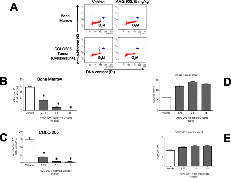
Figure 1
Figure 1
AMG 900 inhibits p-Histone H3 and increases the percentage of G 2 M cells in a dose-dependent manner in COLO 205 tumors and mouse bone marrow measured by Flow Cytometry (FCM). Mice bearing established tumors were orally administered a single dose of vehicle alone or AMG 900 at 3.75, 7.5, or 15 mg/kg. Bone marrow and tumor specimens were collected three hours after treatment (n = 10 per treatment group) and processed for p-Histone H3 and DNA content analysis by FCM. (A) Representative cell cycle profiles of bone marrow (upper panel) and tumor (lower panel, COLO 205 tumor cells were identified using an anti-cytokeratin antibody). AMG 900 treatment decreases the p-Histone H3 positive cell population in G2M detectable in the vehicle-treated control (blue, arrow). Column graphs represent the percentage of p-Histone H3 positive G2M cells (B and C) and G2M cells (D and E) for each treatment group (mean + SE). Statistical significance was determined by comparing the individual AMG 900 treatment groups with vehicle-treated control (*P <0.0001).
Figure 2
Figure 2
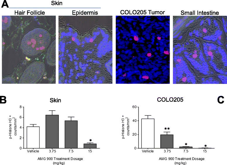
AMG 900 inhibits p-Histone H3 in COLO 205 tumors and mouse skin (hair follicle, epidermis) in a dose-dependent manner measured by Laser Scanning Cytometry (LSC). Mice bearing established tumors were orally administered a single dose of vehicle alone or AMG 900 (as described in Figure 1). Mouse skin specimens (a right- and left-flank punch biopsy from each animal) were collected three hours after treatment (n = 10 per treatment group) and processed for p-Histone H3 analysis by LSC. Small intestine was also collected as a positive staining control for p-Histone H3. Images were captured on a LSC (iCys, CompuCyte) using a 20× objective. See Materials and Methods Section for tissue and tumor section immunofluorescence staining procedure and Additional file 2 Figure S2A for the random sampling contour strategy. (A) Representative scan fields of mouse tissues (skin, hair follicle, small intestine) and COLO 205 tumor showing p-Histone H3 positive objects (red or pink). Nuclei were counterstained with Hoechst 33342 (blue). An average p-Histone H3 object count (per mm2) was determined from three scan fields for each tissue section. Column graphs represent p-Histone H3 positive object for skin (B) and COLO 205 tumor (C) (mean + SE). Statistically significant inhibition of p-Histone H3 compared with vehicle-treated control (*P <0.0001; **P =0.0005).
Figure 3
Figure 3
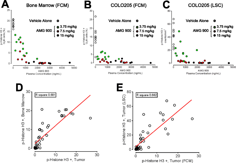
Mouse plasma concentration of AMG 900 correlates with the degree of p-Histone H3 suppression in both COLO 205 tumor and mouse bone marrow tissues. Mice bearing established tumors were orally administered a single dose of vehicle alone or AMG 900 (as described in Figure 1). Mouse plasma was collected three hours after treatment (n = 10 per treatment group). Scatter plots were generated using the observed percentage of p-Histone H3 positive G2M cells for bone marrow (A) and COLO 205 tumor (B and C) and plasma concentration of AMG 900 (ng/mL) from individual mice. Treatment groups are indicated (○ vehicle, “green circle symbol” 3.75, “red circle symbol” 7.5, and ● 15 mg/kg). Regression analysis was used to assess linearity of p-Histone H3 response relationship across tissue sets (D) FCM bone marrow versus tumor, (E) FCM tumor versus LSC tumor. The correlation coefficient (R-squared value) and fitted line (red) are shown for each plot.
Figure 4
Figure 4
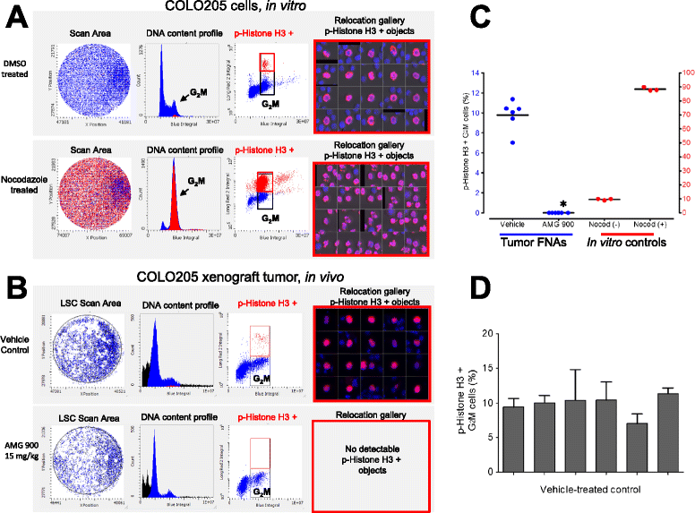
AMG 900 inhibits p-Histone H3 in COLO 205 tumor cells using fine-needle aspirate (FNA) biopsies. (A) LSC based in vitro cell cycle assessment of COLO 205 tumor cells treated with DMSO or 100 ng/mL nocodazole. Cytospin deposited cells were immunostained with an anti-p-Histone H3 antibody and counterstained with DAPI. Plots represent the cell cycle profile indicating G2M (arrow), and p-Histone H3 (red) subpopulations. Relocation gallery of representative scanned images of p-Histone H3 positive cells in mitosis. (B and C) Mice bearing established COLO 205 tumors were orally administered a single dose of vehicle alone or AMG 900 at 15 mg/kg. Tumor FNA punch biopsies (n = 3 per tumor) were collected three hours after treatment (n = 6 per treatment group) and processed for p-Histone H3 and DNA content analysis by LSC. (B) Representative tumor FNA cell cycle profiles of vehicle (upper panel) and AMG 900 (lower panel) treatment groups. AMG 900 treatment completely abolished the p-Histone H3 positive cell population in G2M detectable in the vehicle-treated control (red). (C) Scatter plot represents the individual and mean percentage of p-Histone H3 positive G2M cells for each treatment group. Statistically significant inhibition of p-Histone H3 compared with vehicle-treated control (*P <0.0001). COLO 205 tumor cells were treated with 100 ng/mL nocodazole (Nocod +) or DMSO (Nocod -) as p-Histone H3 staining controls. (D) Column graph represents vehicle-treated control group inter-(6 tumors) and intra-tumor FNA (3 per tumor) variation in the percentage of p-Histone H3 positive G2M cells (mean + SE).
Figure 5
Figure 5
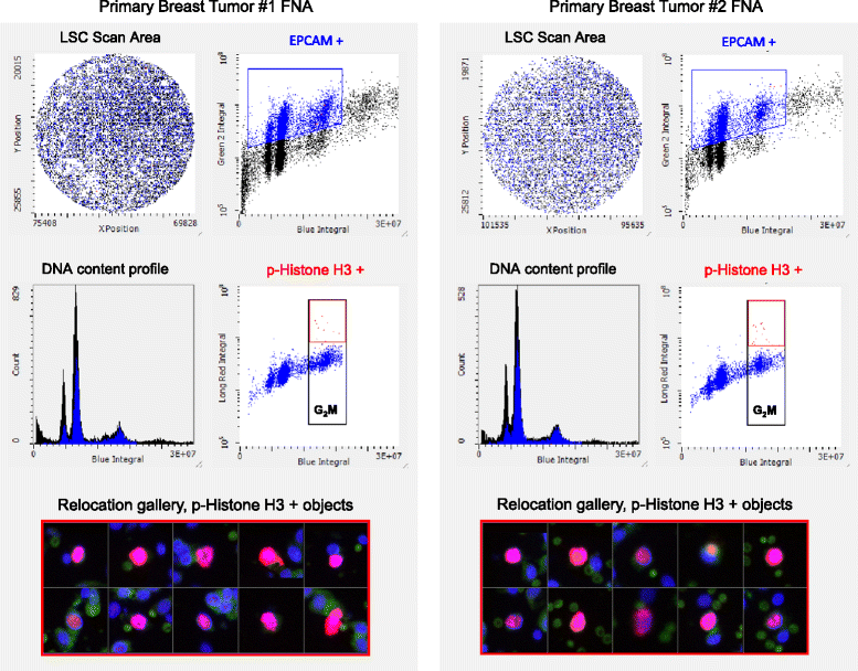
Detection of p-Histone H3 using primary human breast tumor FNA biopsies by LSC. Two mock FNA punch biopsies were collected from the same fresh primary breast tumor tissue. Cytospin deposited cells were immunostained with anti-EpCAM antibody (epithelial cell specific marker) and anti-p-Histone H3 antibodies and counterstained with DAPI. Representative cell cycle profiles of two FNAs showing EpCAM positive (blue) tumor cells with a subpopulation of p-Histone H3 positive G2M cells (red). Relocation gallery showing p-Histone H3 positive objects.

Similar articles

Cited by

References

    1. Warner SL, Bearss DJ, Han H, Von Hoff DD. Targeting aurora-2 kinase in cancer. Mol Cancer Ther. 2003;2:589–595. doi: 10.4161/cbt.2.5.506. - DOI - PubMed
    1. Gizatullin F, Yao Y, Kung V, Harding MW, Loda M, Shapiro GI. The aurora kinase inhibitor VX-680 induces endoreduplication and apoptosis preferentially in cells with compromised p53-dependent postmitiotic checkpoint function. Cancer Res. 2006;66:7668–7677. doi: 10.1158/0008-5472.CAN-05-3353. - DOI - PubMed
    1. Carmena M, Earnshaw WC. The cellular geography of aurora kinases. Nat Rev Mol Cell Biol. 2003;4:842–854. doi: 10.1038/nrm1245. - DOI - PubMed
    1. Jackson JR, Patrick DR, Dar MM, Huang PS. Targeting anti-mitotic therapies: can we improve on tubulin agents? Nat Rev Cancer. 2007;7:107–117. doi: 10.1038/nrc2049. - DOI - PubMed
    1. Harrington EA, Bebbington D, Moore J, Rasmussen RK, Ajose-Adeogun AO, Nakayama T, Graham JA, Demur C, Hercend T, Diu-Hercend A, Su M, Golec JMC, Miller KM. VX-680, a potent and selective small-molecule inhibitor of the aurora kinases, suppresses tumor growth in vivo. Nat Med. 2004;10:262–267. doi: 10.1038/nm1003. - DOI - PubMed

Substances

LinkOut - more resources