. 2015 Feb;25(2):226-34.
doi: 10.1101/gr.179770.114. Epub 2014 Nov 3.

Violation of the 12/23 rule of genomic V(D)J recombination is common in lymphocytes

Affiliations

Violation of the 12/23 rule of genomic V(D)J recombination is common in lymphocytes

Nicholas J Parkinson et al. Genome Res. 2015 Feb.

Abstract

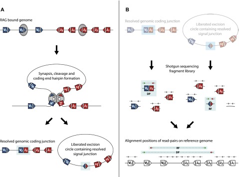
V(D)J genomic recombination joins single gene segments to encode an extensive repertoire of antigen receptor specificities in T and B lymphocytes. This process initiates with double-stranded breaks adjacent to conserved recombination signal sequences that contain either 12- or 23-nucleotide spacer regions. Only recombination between signal sequences with unequal spacers results in productive coding genes, a phenomenon known as the "12/23 rule." Here we present two novel genomic tools that allow the capture and analysis of immune locus rearrangements from whole thymic and splenic tissues using second-generation sequencing. Further, we provide strong evidence that the 12/23 rule of genomic recombination is frequently violated under physiological conditions, resulting in unanticipated hybrid recombinations in ∼10% of Tcra excision circles. Hence, we demonstrate that strict adherence to the 12/23 rule is intrinsic neither to recombination signal sequences nor to the catalytic process of recombination and propose that nonclassical excision circles are liberated during the formation of antigen receptor diversity.


Figures

Figure 1.
Figure 1.
Schematic representation of RAG-mediated V(D)J recombination showing the relative mapping positions of DP and RF read-pairs from recombined genomic and excised circular DNA. (A) Hypothetical genomic locus containing variable (blue box) and joining (red box) elements flanked by recombination signal sequence (RSS) motifs (blue and red triangles) are bound and brought into close association by the recombination activating gene (RAG) complex (gray). RAG-mediated double-stranded cleavage at RSS sites precedes genomic deletion (coding) end processing by the nonhomologous end joining (NHEJ) complex. Coding ends are covalently hair-pinned (gray circle section) and reopened prior to ligation. Secondarily, the excision circle (EC; signal) junction is ligated from unprocessed DNA ends with little modification. (B) Sequencing library fragments from recombined EC and genomic (g) DNA are shown. Read-pair sequences from fragments are mapped back to the reference genome, allowing junction-spanning reads to be identified. Gray arrows represent “standard” read-pairs generated from EC DNA or gDNA and map to the reference genome in standard orientation separated by a mapping distance equal to the initial fragment length. Deletion read-pairs (DPs) spanning a coding junction map to the reference genome with standard relative orientation—the forward read (green) mapping 5′ of and facing the reverse read (red)—but are separated by mapping distances greater than the initial fragment length. Reverse-forward read-pairs (RFs) spanning the signal junction map to the reference genome in an inverted relative read orientation so that the reverse read (red) maps 5′ of and faces away from the forward read (green). RF read-pairs are separated by the full length of the circle from which they originate rather than the initial library fragment length.
Figure 2.
Figure 2.
EC-seq enriches Tcra locus-associated circularized excision material. (A) Gross coverage of perfectly aligned read-pairs (AA) across the mouse Tcra locus on Chromosome 14 in whole thymus-captured EC-seq material. (B) Meta-analysis of Vα-Jα RF read-pairs (top) or DP read-pairs (bottom) in EC-seq libraries showing close association of reads with known Vα element subregion RSSs (left) or Jα element subregion RSSs (right). The data set comprises thymic and splenic EC-seq replicates. Overlay of forward reads is shown in green; overlay of reverse reads is shown in red. Broken vertical lines indicate the RSS cleavage site. (C) Heatmap analysis of 31,535 RSS-associated Vα-Jα RF junctions from EC-seq metadata displaying the diversity of recombined Vα-Jα ECs. Data are from three thymic and three splenic replicates. ECs are only shown for active elements with uniquely mappable sequences. (D) EC-seq pile-up of Jα-Jα RF read-pairs across the Jα segment subregion showing clustering of Jα element RSSs. Forward reads are shown in green; reverse reads are shown in red. (E) Meta-analysis of Jα-Jα subregion RFs relative to known Jα region RSS sites. Forward reads are shown in green; reverse reads are shown in red. (F) Ratio of Jα-Jα (blue bars) and Vα-Vα (red bars) to Vα-Jα RFs in three thymic and three splenic EC-seq libraries.
Figure 3.
Figure 3.
Jα-Jα ECs occur predominantly with their nearest neighbor. (A) Graphical representation of individual Jα-Jα ECs from a whole thymus EC-seq library showing connectivity and relative frequency of Jα-Jα ECs across the Jα element subregion. Ribbon width represents the frequency of recombinations between connected segments. Ribbon color is alternated to discriminate between independent ribbon connections at the same segment. (B) Heatmap meta-analysis of 33,332 Jα-Jα RFs showing the frequency of individual EC events. (C) Jα-Jα EC events from thymic and splenic EC-seq libraries showing the frequency of excisions spanning neighboring or multiple genomic Jα segments.
Figure 4.
Figure 4.
IR-seq analysis of T cell developmental stages confirms that Jα-Jα ECs originate during alpha chain rearrangement. (A) Schematic diagram of T cell development showing stage-specific, thymus-derived cell sorts used to generate 12 IR-seq libraries (see Methods). (B) Coverage in the intermediate CD4 single positive (ISP) IR-seq library across the Tcra locus. Coverage was computed between neighboring Vα, Dα, and Jα gene segment RSSs and normalized relative to coverage in the same regions in the prerecombinational DN1 IR-seq data set. Depleted coverage across the Tcr delta locus is predicted to result from genomic excision events and loss of delta ECs during preceding proliferative phases. Coverage across the Jα subregion remains at ∼1, indicating that Jα-Jα ECs are not overrepresented in IR-seq. (C) Heatmap analyses of sorted T cell precursors showing Vα-Jα EC events (top) and Jα-Jα EC events (bottom) for eight consecutive recombinationally active IR-seq libraries. (D) Jα-Jα–to–Vα-Jα (blue bars) and Vα-Vα–to–Vα-Jα (red bars) ratios of RFs in 12 IR-seq libraries, representing key stages of T cell development. (E) DP-to-RF ratios for Vα-Jα (blue bars) and Jα-Jα (red bars) in 12 IR-seq libraries representing key stages of T cell development.
Figure 5.
Figure 5.
Analysis of Vα-Jα coding, Vα-Jα signal, and Jα-Jα hybrid junctions shows that Jα-Jα hybrids have processing characteristics similar to Vα-Jα coding junctions. (A) Frequency of nucleotide deletions flanking the RSS site of cleavage in coding Vα-Jα junctions (blue line), signal Vα-Jα junctions (black line), and hybrid Jα-Jα junctions (purple line). (B) Length of nontemplated bases in coding Vα-Jα junctions (blue line), signal Vα-Jα junctions (black line), and hybrid Jα-Jα junctions (purple line).
Figure 6.
Figure 6.
Alternative models of J-J EC production. (A) V2-J3 recombination (see Fig. 1) forms an excision circle pre-ligation intermediate containing variable (blue box) and joining (red box) gene segments with RAG complexes (gray circles) bound at multiple RSS sites (red and blue triangles). Additional synapsis and dsDNA cleavage occurs between an internal J segment RSS and RAG complex bound to either unresolved signal ends or a fully ligated signal junction (as denoted by a dashed line). V-J signal and J-J hybrid junction ends (filled circles) are resolved and liberated as independent ECs. (B) V2-J3 recombination forms a coding junction pre-ligation intermediate with hair-pinned ends (gray circles) containing variable (blue box) and joining (red box) gene segments with RAG complexes (gray circles) bound at multiple RSS sites (red and blue triangles). Additional synapsis and dsDNA cleavage occurs between the RAG complex bound to the unresolved coding end and the flanking J2 segment RSS. The initial V2-J3 coding junction is “skipped,” and a V2-J2 coding junction is formed along with a J-J hybrid junction containing EC.

Similar articles

Cited by

References

    1. Alexandre D, Chuchana P, Roncarolo MG, Yssel H, Spits H, Lefranc G, Lefranc MP. 1991. Reciprocal hybrid joints demonstrate successive V-J rearrangements on the same chromosome in the human TCRγ locus. Int Immunol 3: 973–982. - PubMed
    1. Bogue MA, Wang C, Zhu C, Roth DB. 1997. V(D)J recombination in Ku86-deficient mice: distinct effects on coding, signal, and hybrid joint formation. Immunity 7: 37–47. - PubMed
    1. Bredemeyer AL, Sharma GG, Huang CY, Helmink BA, Walker LM, Khor KC, Nuskey B, Sullivan KE, Pandita TK, Bassing CH, et al. . 2006. ATM stabilizes DNA double-strand-break complexes during V(D)J recombination. Nature 442: 466–470. - PubMed
    1. Briney BS, Willis JR, Hicar MD, Thomas JW, Crowe JE Jr. 2012. Frequency and genetic characterization of V(DD)J recombinants in the human peripheral blood antibody repertoire. Immunology 137: 56–64. - PMC - PubMed
    1. Carroll AM, Slack JK, Mu X. 1993. V(D)J recombination generates a high frequency of nonstandard TCR Dδ-associated rearrangements in thymocytes. J Immunol 150: 2222–2230. - PubMed

Publication types