. 2015 Jan;129(1):53-64.
doi: 10.1007/s00401-014-1358-z. Epub 2014 Nov 4.

Hippocampal sclerosis in Lewy body disease is a TDP-43 proteinopathy similar to FTLD-TDP Type A

Affiliations

Hippocampal sclerosis in Lewy body disease is a TDP-43 proteinopathy similar to FTLD-TDP Type A

Naoya Aoki et al. Acta Neuropathol. 2015 Jan.

Abstract

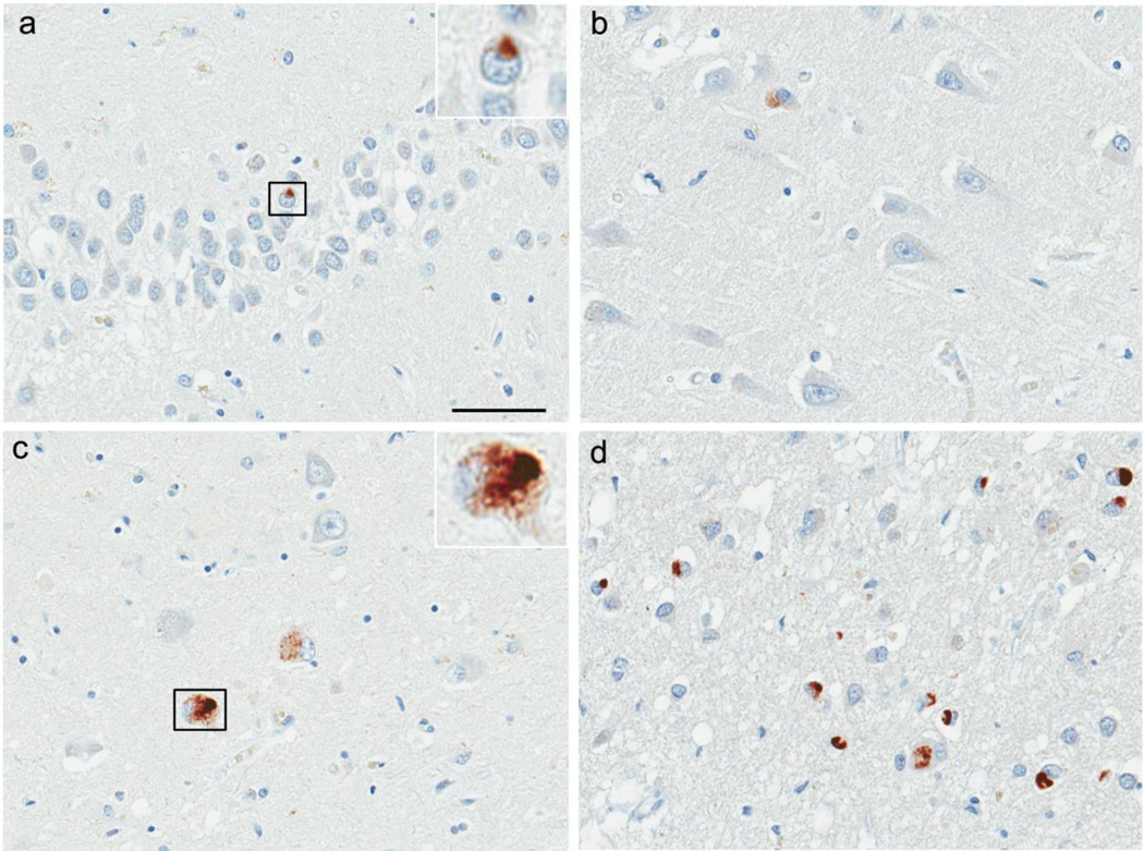
Hippocampal sclerosis (HpScl) is frequent in frontotemporal lobar degeneration with TDP-43 pathology (FTLD-TDP), but it also occurs in dementia of the elderly with or without accompanying Alzheimer type pathology. HpScl has been hypothesized to be a neurodegenerative process given its association with TDP-43 pathology, but this is still controversial. TDP-43 pathology is found in Lewy body disease (LBD), but no study has focused on the pathologic and genetic characteristics of HpScl in LBD. We found HpScl in 5.2% of 669 LBD cases (289 transitional and 380 diffuse). Older age, higher Braak neurofibrillary tangle (NFT) stage, and presence of TDP-43 pathology were associated with HpScl. There was no difference in the frequency of HpScl between transitional and diffuse LBD, suggesting that Lewy-related pathology appears to have no direct association with HpScl. All HpScl cases had TDP-43 pathology consistent with Type A pattern. HpScl cases harbored genetic variation in TMEM106B that has been previously associated with FTLD-TDP. Interestingly, the severity of TDP-43-positive fine neurites in CA1 sector, a possible pathologic precursor of HpScl, was associated with the TMEM106B variant. These results demonstrate HpScl in LBD is a TDP-43 proteinopathy and is similar to FTLD-TDP Type A. Furthermore, a subset of LBD cases without HpScl ("pre-HpScl") had similar pathologic and genetic characteristics to typical HpScl, suggesting that the spectrum of HpScl pathology may be wider than previously thought. Some cases with many extracellular NFTs also had a similar profile. We suggest that HpScl is "masked" in these cases.


Figures

Fig. 1
Fig. 1
Immunohistochemistry for TDP-43 in a hippocampal sclerosis case (Type A). Neuronal cytoplasmic inclusions (NCIs) in the dentate fascia (a). Fine neurites in the CA1 sector (b, c) and subiculum (d). Mixture of NCIs and dystrophic neurites (DNs) in the parahippocampal gyrus (e), inferior temporal gyrus (f), and amygdala (g). Inset in (f) shows a neuronal intranuclear inclusion with a lentiform shape. Sparse TDP-43 pathology in the putamen (h) and substantia nigra (i). Insets in (h) and (i) show a NCI and a DN, respectively. TDP-43 pS409/410 (a–i). Scale bars: a, b, d–I; 50um, C; 200um.
Fig. 2
Fig. 2
Immunohistochemistry for TDP-43 in the case without hippocampal sclerosis (Type B/NFT). Sparse neuronal cytoplasmic inclusions (NCIs) in the dentate fascia (a). Inset in (a) shows a NCI. TDP-43-positive fine neurites are not present in the CA1 sector (b) and subiculum (c). TDP-43 immunoreactivity is shown in some NFTs in the subiculum (c). Inset in (c) shows a NFT-like TDP-43 pathology. Mixture of NCIs and NFT-like TDP-43 pathology in the parahippocampal gyrus with sparse dystrophic neurites (d). TDP-43 pS409/410 (a–d). Scale bars: a–d; 50µm.
Fig. 3
Fig. 3
TDP-43-positive fine neurites in CA1 (a, b) were similar in HpScl-Type A (a) and Not HpScl-Type A High CA1 neurites (b) compared to Not HpScl-Type A Low CA1 neurites (c). CA1 fine neurites are significantly less in all Type A cases (a–c) with TMEM106B CC genotypes than in cases with TT (p=0.002) or CT genotype (p=0.018) (d). Not HpScl-Type A cases with High CA1 fine neurites had low frequency of TMEM106B CC genotype similar to HpScl-Type A cases (e). The boxes show median and 25th and 75th percentiles with whisker plots showing 10th and 90th percentiles. Scale bars: a–c; 50µm.
Fig. 4
Fig. 4
We propose a classification of hippocampal sclerosis in LBD based upon neuronal loss, TDP-43 pathology and extracellular NFTs in the CA1 sector (a). “Typical-HpScl “ has neuronal loss and TDP-43-positive fine neurites but no or relatively mild NFTs when compared with the degree of neuronal loss (b, e, h). “Pre-HpScl” has none to minimal neuronal loss or extracellular NFTs, but abundant TDP-43-positive fine neurites (c, f, i). “Masked HpScl” has neuronal loss and many extracellular NFTs but also abundant TDP-43-positive fine neurites (d, g, j). Arrows in (d) show extracellular NFTs. Hematoxylin & eosin (b–d), TDP-43 pS409/410 (e–g), Tau PHF-1 (h–j). Scale bars: b–j; 50um. NL=neuronal loss, eNFTs=extracellular neurofibrillary tangles, H&E=hematoxylin & eosin.

References

    1. Ala TA, Beh GO, Frey WH., 2nd Pure hippocampal sclerosis: a rare cause of dementia mimicking Alzheimer's disease. Neurology. 2000;54:843–848. - PubMed
    1. Amador-Ortiz C, Lin WL, Ahmed Z, et al. TDP-43 immunoreactivity in hippocampal sclerosis and Alzheimer's disease. Ann Neurol. 2007;61:435–445. - PMC - PubMed
    1. Arai T, Mackenzie IR, Hasegawa M, et al. Phosphorylated TDP-43 in Alzheimer's disease and dementia with Lewy bodies. Acta Neuropathologica. 2009;117:125–136. - PubMed
    1. Braak H, Braak E. Neuropathological stageing of Alzheimer-related changes. Acta Neuropathol. 1991;82:239–259. - PubMed
    1. Dickson DW, Baker M, Rademakers R. Common variant in GRN is a genetic risk factor for hippocampal sclerosis in the elderly. Neuro-degenerative diseases. 2010;7:170–174. - PMC - PubMed

Publication types

MeSH terms