. 2014 Nov 4:12:309.
doi: 10.1186/s12967-014-0309-8.

Specific-detection of clinical samples, systematic functional investigations, and transcriptome analysis reveals that splice variant MUC4/Y contributes to the malignant progression of pancreatic cancer by triggering malignancy-related positive feedback loops signaling

Affiliations

Specific-detection of clinical samples, systematic functional investigations, and transcriptome analysis reveals that splice variant MUC4/Y contributes to the malignant progression of pancreatic cancer by triggering malignancy-related positive feedback loops signaling

Yi Zhu et al. J Transl Med. .

Abstract

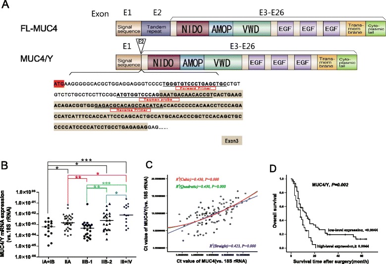
Background: MUC4 plays important roles in the malignant progression of human pancreatic cancer. But the huge length of MUC4 gene fragment restricts its functional and mechanism research. As one of its splice variants, MUC4/Y with coding sequence is most similar to that of the full-length MUC4 (FL-MUC4), together with alternative splicing of the MUC4 transcript has been observed in pancreatic carcinomas but not in normal pancreas. So we speculated that MUC4/Y might be involved in malignant progression similarly to FL-MUC4, and as a research model of MUC4 in pancreatic cancer. The conjecture was confirmed in the present study.

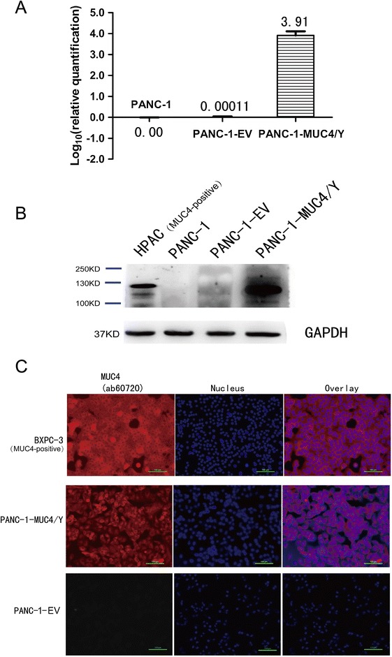
Methods: MUC4/Y expression was detected by real-time quantitative reverse transcription polymerase chain reaction (qRT-PCR) using gene-specific probe in the clinic samples. The effects of MUC4/Y were observed by serial in vitro and in vivo experiments based on stable over-expressed cell model. The underlying mechanisms were investigated by sequence-based transcriptome analysis and verified by qRT-PCR, Western blot and enzyme-linked immunosorbent assays.

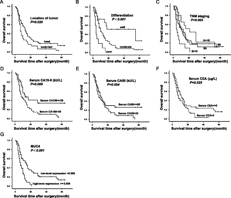
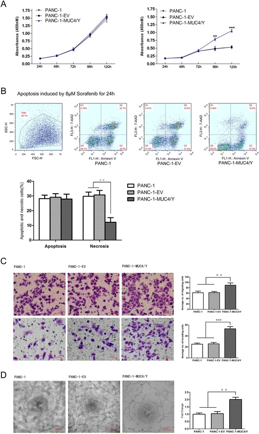
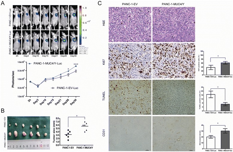
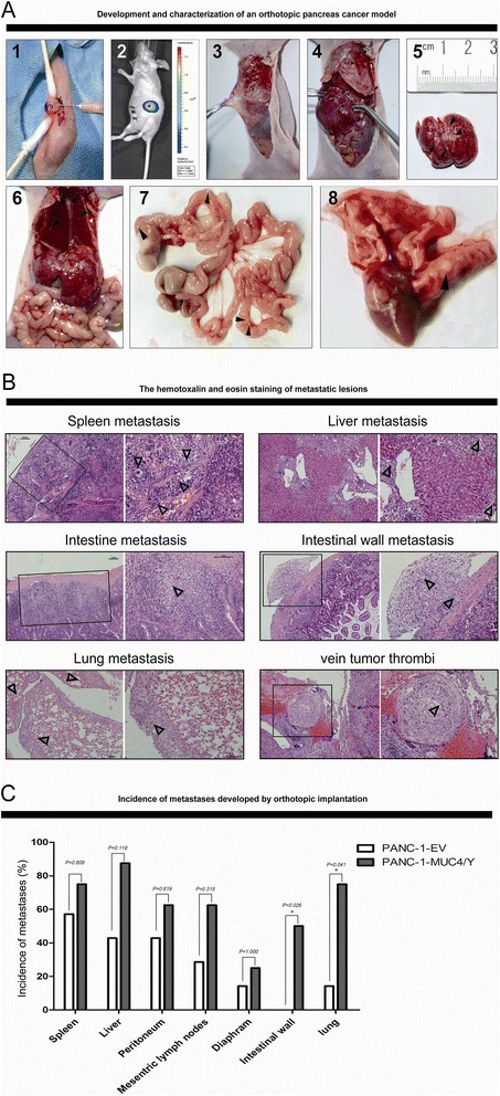
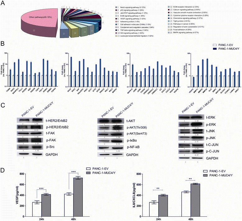
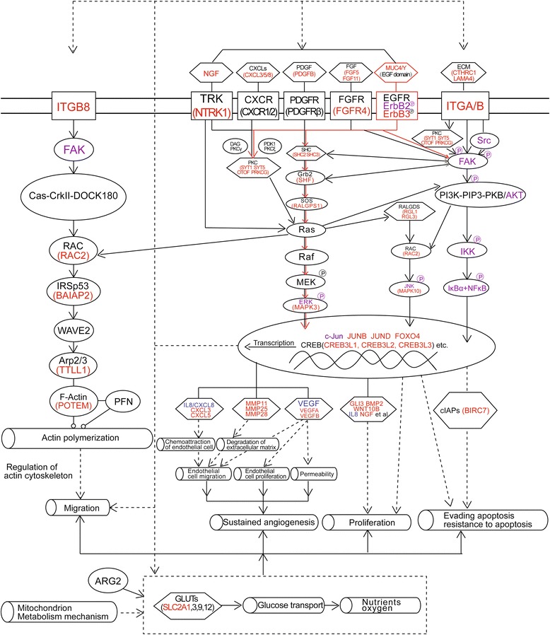
Results: The detection of clinical samples indicates that MUC4/Y is significantly positive-correlated with tumor invasion and distant metastases. Based on stable forced-expressed pancreatic cancer PANC-1 cell model, functional studies show that MUC4/Y enhances malignant activity in vitro and in vivo, including proliferation under low-nutritional-pressure, resistance to apoptosis, motility, invasiveness, angiogenesis, and distant metastasis. Mechanism studies indicate the novel finding that MUC4/Y triggers malignancy-related positive feedback loops for concomitantly up-regulating the expression of survival factors to resist adverse microenvironment and increasing the expression of an array of cytokines and adhesion molecules to affect the tumor milieu.

Conclusions: In light of the enormity of the potential regulatory circuitry in cancer afforded by MUC4 and/or MUC4/Y, repressing MUC4 transcription, inhibiting post-transcriptional regulation, including alternative splicing, or blocking various pathways simultaneously may be helpful for controlling malignant progression. MUC4/Y- expression model is proven to a valuable tool for the further dissection of MUC4-mediated functions and mechanisms.


Figures

Figure 1
Figure 1
Significant positive correlation between MUC4/Y mRNA expression level and TNM stage, MUC4 mRNA expression level, and survival in PDAC. (A) Schematic representation of the design strategy for specific primers and TaqMan probe in MUC4/Y gene detection based on the difference between the exon sequences of MUC4/Y (NCBI Reference Sequence: NM_004532.5) and FL-MUC4 (NCBI Reference Sequence: NM_018406.6). The TaqMan probe sequence lies in the exon 1–exon 3 junction; as exon 2 is absent, it detects MUC4/Y expression rather than other MUC4 types encoding the exon 1–exon 2 junction, or MUC4/X (NCBI Reference Sequence: NM_138297.4), which encodes the exon 1–exon 4 junction because it lacks the coding exons 2 and 3. Primers and TaqMan probe are underlined. (B) Comparison of MUC4/Y mRNA expression at different TNM stages. Scatter dot plots were drawn from the minimum extending to the maximum; the center horizontal line denotes the sample median. *P <0.05, **P ≤0.01, ***P ≤0.001. (C) Positive correlation between MUC4/Y and MUC4 mRNA expression (R 2 = 0.430, P <0.001) and curve fitting. Regression equations were Y = 6.553 + 0.3212 × X + 0.02719 × X/2–0.00033333 × X/3 (cubic), Y = 6.522 + 0.3514 × X + 0.02102 × X/2 (quadratic), and Y = 5.915 + 0.6127 × X (straight line). According to the correlation coefficient value, the cubic or quadratic curve model was the better model. Ct, threshold cycle value. (D) Kaplan–Meier survival curves of PDAC patients according to MUC4/Y mRNA expression status. The P-value was calculated using the log-rank test.
Figure 2
Figure 2
Kaplan-Meier survival estimate by clinicopathological factors for 108 PDAC patients who underwent pancreaticoduodenectomy and survived at least 30 days after surgery. The p value was calculated by the Log-rank test. The correlation between survival and tumor location (A), tumor differentiation (B), TNM staging (C), serum level of CA19-9 (D), serum level of CA50 (E), serum level of CEA (F), MUC4 mRNA expression status (G), respectively, among which the P value of serum level of CA50 lied in the critical (P = 0.054).
Figure 3
Figure 3
MUC4/Y expression and subcellular localization in PANC-1 cells. (A) Real-time PCR using specific primers and TaqMan probe to examine MUC4/Y transcript expression in PANC-1-EV cells and PANC-1-MUC4/Y cells. The level of target gene expression in the PANC-1-MUC4/Y cells was 9912-fold and 9808-fold higher than that of the blank control and negative control, respectively. (B) Western blot confirmation of MUC4/Y protein expression. Total protein from cell extracts was resolved on precast gels. The signal was detected using an electrochemiluminescence reagent kit. (C) Immunofluorescence demonstrating MUC4/Y subcellular localization similar to that of wild-type MUC4. The pancreatic cancer cell lines of HPAC and BXPC-3 is MUC4 positive-expression as positive control for specific antibody.
Figure 4
Figure 4
MUC4/Y enhances PANC-1 cell malignant activity in vitro . Data from three repeated experiments are presented as means ± SD. **P <0.01, ***P <0.001 vs. controls. (A) MUC4/Y enhanced in vitro proliferation of PANC-1 cells under stress from low nutritional status. The absorbance values of cells at different time points were detected with WST-8 dye. Cells were maintained in medium containing 10% serum (left) and 1% serum (right). (B) MUC4/Y increased resistance to apoptotic reagents, i.e., sorafenib. Representative templates of FACS analysis showing the proportion of cells positive for annexin V and 7-AAD (top right quadrant) representing the percentage of necrotic cells; the proportion of cells that were annexin V–positive and 7-AAD–negative (bottom right quadrant) represented the percentage of apoptotic cells (top). Bar denotes the percentage of apoptotic and necrotic cells in PANC-1–derived clones (bottom). (C) MUC4/Y affected pancreatic cancer cell metastatic potential in vitro. Bar graph shows the number of PANC-1–derived clones that had migrated or invaded through the Matrigel. (D) MUC4/Y enhanced cancer cell–associated HUVEC endotube formation. Bar denotes the fold increase of the number of endotubes compared to the blank control.
Figure 5
Figure 5
MUC4/Y contributed to increase tumor growth with rising proliferative activity and MVD and decreased apoptosis in vivo. (A) In vivo BLI showing the tumor growth rates over time in the subcutaneous model. Up: Representative luminescence images for each group. Down: Tumor growth rates indicated by bioluminescence (photons/s) in PANC-1-MUC4/Y-Luc (n = 6) and control (n = 6) BALB/c nude mice at 2 hours and 3, 10, 15, 21, 26, 30 days after tumor cell injection; bars, SE. *P <0.05, ***P <0.001. (B) Subcutaneous tumors and their size (mm) from two groups measured at the 30-day time point when mice were sacrificed. Scatter dot, tumor size of every mouse; the center horizontal line, mean. *P <0.05. (C) Histological and IHC analysis of subcutaneous tumors. Histologically, there was no difference between subcutaneous tumors of the PANC-1-MUC4/Y-Luc group and control groups (H&E staining, ×400 magnification). Ki67, TUNEL, and CD31 staining is of paraffin-embedded sections from solid tumors. Micrographs are representative images of two groups (Ki67, TUNEL, CD31: ×400, 200, 100 magnification, respectively). Charts depict the mean proportion of Ki67- and TUNEL-positive cells and the average number of CD31-positive microvessels per field, respectively. Five fields per slide and at least five slides per group were examined and compared using the Student t-test. Columns, mean; bars, SD; *P <0.05.
Figure 6
Figure 6
Development of orthotopic pancreatic cancer model for observation of metastasis. (A) Development of orthotopic pancreatic cancer and gross inspection. 1. PANC-1–derived clones were orthotopically implanted in the parenchyma of the pancreatic head. 2. Luciferase activity imaging at two days following tumor cell injection confirming orthotopic implantation at the injection site without cell spillage. 3–8. Metastasis to the peritoneum, peritoneum and liver, liver, diaphragm and liver, mesenteric lymph node and intestinal wall, and lung, indicated by arrowheads (▲), were obvious macroscopically. (B) Histological analysis of metastatic lesions. Metastatic lesions of the spleen, liver, downward mucosa of the intestine, intestinal wall, lung, vein tumor thrombi (H&E staining, left: ×100 magnification; right: ×200 magnification, Δ indicates tumor). (C) Increased incidences of metastasis at different sites in MUC4/Y overexpression group compared to the control group (n = 8 per group). Comparisons between groups were tested with the chi-square test, *P <0.05.
Figure 7
Figure 7
qRT-PCR, western blotting, and ELISA verification of DEGs related to the malignant functions of MUC4/Y and VEGF family molecules. (A) Distribution of DEGs in the main pathways related to oncogenic transformation showing the change derived from MUC4/Y overexpression–associated molecular signatures; the proportions of each pathway (represented by different colors) are graphed. The DEG list for annotations is the intersection set of PANC-1-MUC4/Y compared to the controls; absolute value of log2 ratio ≥1. (B) Representative qPCR validation results of 44 DEGs and VEGF family molecules (VEGFA, B, C) in PANC-1-MUC4/Y and control cell. (C) Western blot analysis verified a significant upregulation of t-HER2/ErbB2, p-HER2/ErbB2, t-FAK, p-FAK, p-Src, p-AKT(Thr308), p-AKT(Ser473), p-IκBα, p-NF-kB, t-ERK, p-ERK, t-JNK, p-JNK, and p-C-JUN in PANC-1-MUC4/Y compared with control. (D) ELISA of VEGF and IL8/CXCL8 using cell culture supernatants. Left: VEGF; right: IL8/CXCL8 production from PANC-1–derived clones incubated for 24 h or 48 h. Bars denote mean ± SD, ***P <0.001, **P <0.01.
Figure 8
Figure 8
MUC4/Y-dependent pathways involved in malignant activity. Scheme depicts gene complexes and families (hexagons), membrane receptors (rectangles), function (hollow cylinders), and others (ovals). Red, purple, and blue key factors in the signaling pathways were verified by qRT-PCR, western blotting, and ELISA, respectively. Red lines denote the triggers.

Similar articles

Cited by

References

    1. Moniaux N, Escande F, Porchet N, Aubert JP, Batra SK. Structural organization and classification of the human mucin genes. Front Biosci. 2001;6:D1192–D1206. doi: 10.2741/Moniaux. - DOI - PubMed
    1. Porchet N, Nguyen VC, Dufosse J, Audie JP, Guyonnet-Duperat V, Gross MS, Denis C, Degand P, Bernheim A, Aubert JP. Molecular cloning and chromosomal localization of a novel human tracheo-bronchial mucin cDNA containing tandemly repeated sequences of 48 base pairs. Biochem Biophys Res Commun. 1991;175:414–422. doi: 10.1016/0006-291X(91)91580-6. - DOI - PubMed
    1. Moniaux N, Nollet S, Porchet N, Degand P, Laine A, Aubert JP. Complete sequence of the human mucin MUC4: a putative cell membrane-associated mucin. Biochem J. 1999;338(Pt 2):325–333. doi: 10.1042/0264-6021:3380325. - DOI - PMC - PubMed
    1. Khorrami AM, Choudhury A, Andrianifahanana M, Varshney GC, Bhattacharyya SN, Hollingsworth MA, Kaufman B, Batra SK. Purification and characterization of a human pancreatic adenocarcinoma mucin. J Biochem. 2002;131:21–29. doi: 10.1093/oxfordjournals.jbchem.a003073. - DOI - PubMed
    1. Gross MS, Guyonnet-Duperat V, Porchet N, Bernheim A, Aubert JP, Nguyen VC. Mucin 4 (MUC4) gene: regional assignment (3q29) and RFLP analysis. Ann Genet. 1992;35:21–26. - PubMed

Publication types