Review
. 2015 Jan;359(1):65-85.
doi: 10.1007/s00441-014-2011-9. Epub 2014 Nov 1.

The roles and regulation of Polycomb complexes in neural development

Affiliations
Review

The roles and regulation of Polycomb complexes in neural development

Matthew Corley et al. Cell Tissue Res. 2015 Jan.

Abstract

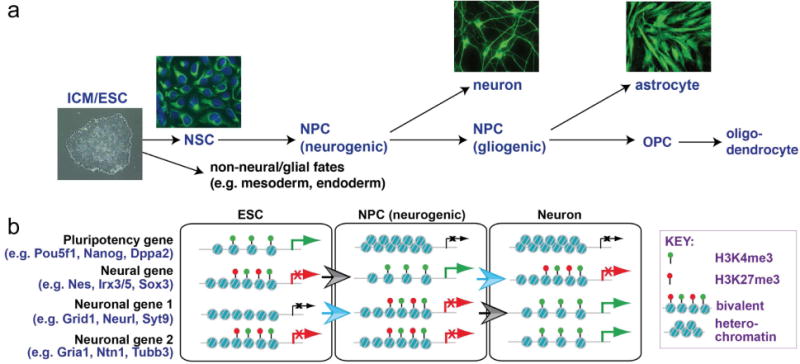
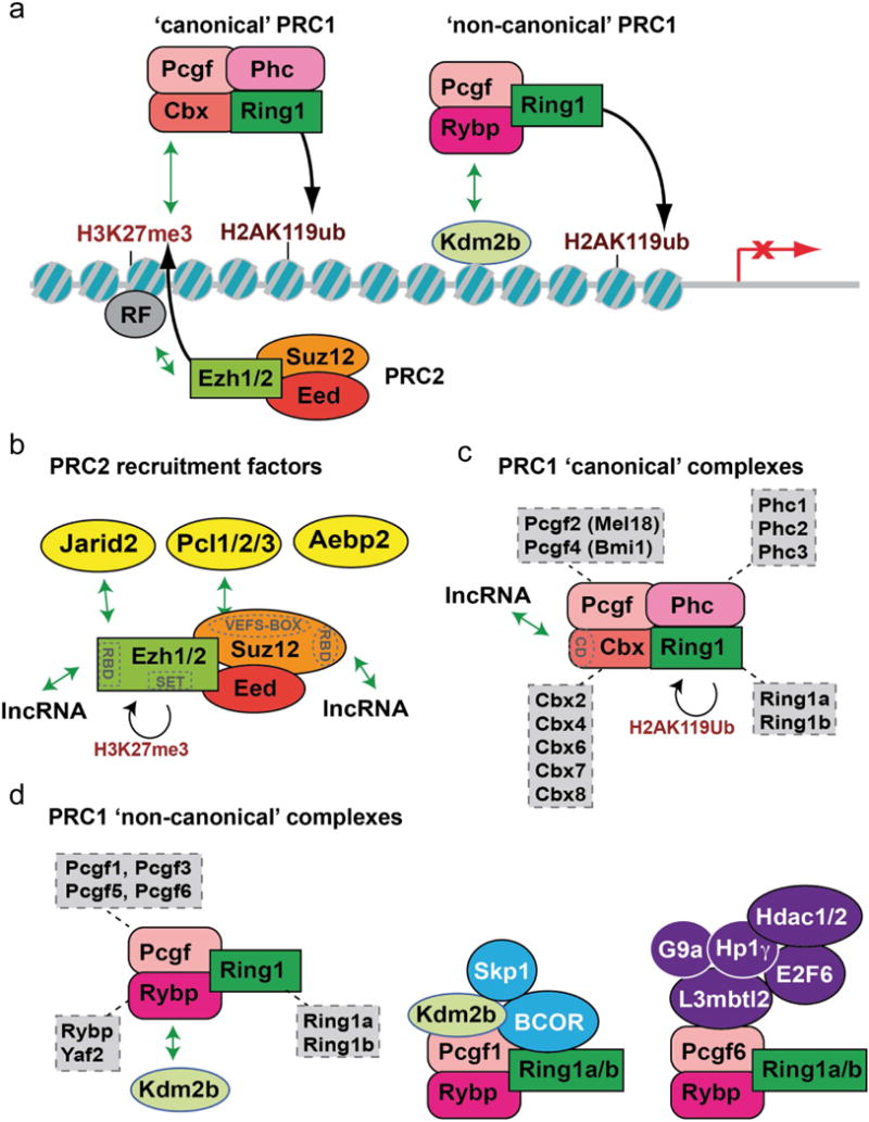
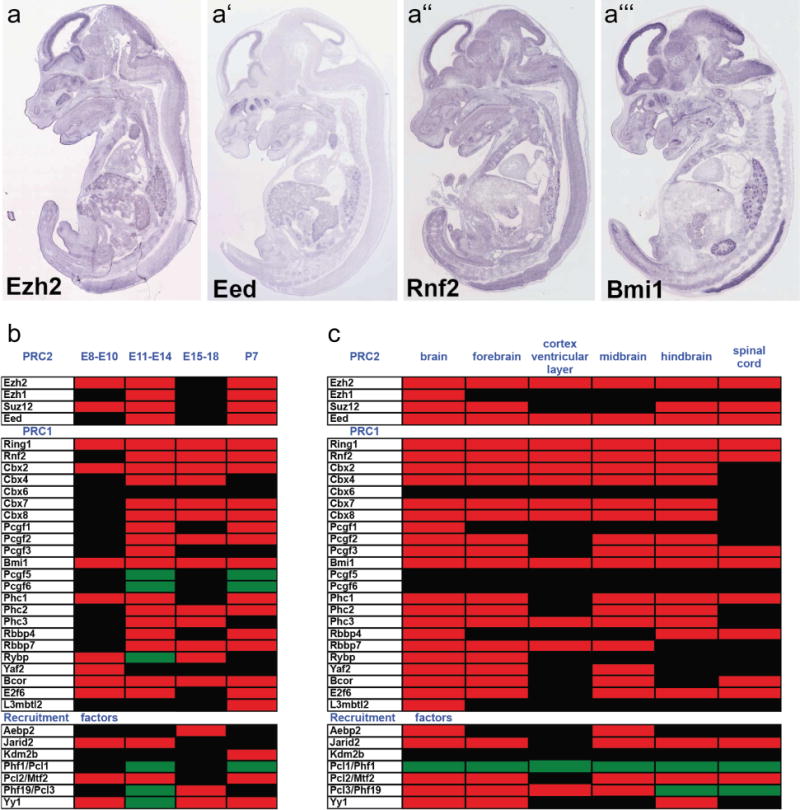
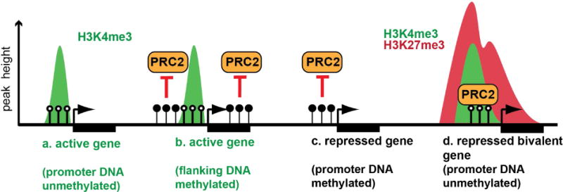
In the developing mammalian nervous system, common progenitors integrate both cell extrinsic and intrinsic regulatory programs to produce distinct neuronal and glial cell types as development proceeds. This spatiotemporal restriction of neural progenitor differentiation is enforced, in part, by the dynamic reorganization of chromatin into repressive domains by Polycomb repressive complexes, effectively limiting the expression of fate-determining genes. Here, we review the distinct roles that Polycomb repressive complexes play during neurogenesis and gliogenesis, while also highlighting recent work describing the molecular mechanisms that govern their dynamic activity in neural development. Further investigation of the way in which Polycomb complexes are regulated in neural development will enable more precise manipulation of neural progenitor differentiation facilitating the efficient generation of specific neuronal and glial cell types for many biological applications.


Figures

Figure 1
Figure 1. Changes in histone modification state accompany cell state transitions during neural development
(a) Pluripotent cells of the early embryo (inner cell mass=ICM) and embryonic stem cells (ESCs) can differentiate into multiple neuronal and glial cell types. This process involves cellular transitions through distinct progenitor cell states. Photographs show human ESCs and their directed differentiation into Nestin-expressing neural stem cells (NSCs: green, with DAPI counterstaining of nuclei), Beta-III-tubulin-expressing neurons, and GFAP-expressing astrocytes. (b) Chromatin modification state changes occur at lineage-specific genes during the transition of pluripotent ESCs into differentiated derivatives including neurons. Hallmarks of this process include acquisition of a bivalent modification state by new sets of genes in each progenitor type. Bivalent genes are silent, but primed for rapid expression as differentiation occurs. As a progenitor cell differentiates, bivalent genes associated with the selected lineage resolve into an active chromatin state, while genes associated with alternative lineages adopt more stable heterochromatic configurations. Blue open arrow demarcates transition to a poised bivalent state and gray arrows demarcate transitions from a bivalent to an active (H3K4me3 only) state. Examples for each type of gene are from Mohn et al., 2008.
Figure 2
Figure 2. Polycomb complex diversity
(a) H3K27me3 modification of chromatin by the EZH1/2 subunit of PRC2 promotes recruitment of the ‘canonical’ PRC1 complex, which catalyzes ubiquitylation of H2AK119 to repress gene expression. Additional ‘non-canonical’ forms of the PRC1 complex can be recruited to chromatin in a PRC2-independent manner. (b) PRC2 associates with several non-stoichiometric proteins, shown in yellow. PRC2 subunits EZH2 and SUZ12 also have functional domains described in text (shown in gray), including RNA binding domains (RBDs) that can associate with long non-coding RNAs (lncRNA). Both protein and lncRNA associations have the potential to facilitate context-dependent recruitment to chromatin. (c) ‘Canonical’ PRC1 complexes have multiple forms, through inclusion of specific subunit variants. The chromodomain (CD) of the Cbx subunit can also associate with lncRNAs. (d) Non-canonical PRC1 complexes have an even more diverse potential subunit composition, with some complex variants shown here (see text for further information).
Figure 3
Figure 3. Expression of Polycomb complex proteins and recruitment factors in the developing nervous system
(a–a′″) In situ hybridization data for four core Polycomb proteins at embryonic day 14.5 (Genepaint database) shows robust expression in the ventricular zone of the cortex and other CNS locations. (b–c) The Gene Expression Database at the Mouse Genome Informatics Resource was used to catalog (b) temporal and (c) regional expression of Polycomb subunits that has been reported for the mouse central nervous system between embryonic day 8 (E8) and post-natal day 7 (P7). Heat maps indicate developmental time windows (b) or CNS regions (c) where expression is documented as present (red), as absent (green), or where no data is reported (black). CNS expression of some Polycomb core subunits is reported from the onset of neural plate formation, with expression of other subunits being detected during neurogenesis and through post-natal stages. Within the CNS, expression of many subunits is reported in the fore-, mid-, and hindbrain regions, with core PcG subunits in particular showing enrichment in the ventricular zone of the cortex. Expression of some subunits has also been reported in the spinal cord.
Figure 4
Figure 4. Relationship between DNA methylation and Polycomb recruitment
(a–b) Promoters of active genes are frequently (a) unmethylated (open circles), although (b) DNA methylation (closed circles) flanking the promoter can also facilitate the active state by preventing PRC2-mediated repression. Repressed genes may have methylated promoter DNA (c), although demethylation allows PRC2-mediated silencing (d). Arrows indicate the transcription start site and rectangles indicate gene bodies.

References

    1. Alder O, Lavial F, Helness A, Brookes E, Pinho S, Chandrashekran A, Arnaud P, Pombo A, O’Neill L, Azuara V. Ring1B and Suv39h1 delineate distinct chromatin states at bivalent genes during early mouse lineage commitment. Development. 2010;137(15):2483–2492. - PMC - PubMed
    1. Aloia L, Di Stefano B, Sessa A, Morey L, Santanach A, Gutierrez A, Cozzuto L, Benitah SA, Graf T, Broccoli V, Di Croce L. Zrf1 is required to establish and maintain neural progenitor identity. Genes Dev. 2014;28(2):182–197. - PMC - PubMed
    1. Aprea J, Prenninger S, Dori M, Ghosh T, Monasor LS, Wessendorf E, Zocher S, Massalini S, Alexopoulou D, Lesche M, Dahl A, Groszer M, Hiller M, Calegari F. Transcriptome sequencing during mouse brain development identifies long non-coding RNAs functionally involved in neurogenic commitment. EMBO J. 2013;32(24):3145–3160. - PMC - PubMed
    1. Armstrong L. Epigenetic control of embryonic stem cell differentiation. Stem Cell Rev. 2012;8(1):67–77. - PubMed
    1. Arney KL, Fisher AG. Epigenetic aspects of differentiation. J Cell Sci. 2004;117(Pt 19):4355–4363. - PubMed

MeSH terms

Substances