Review
. 2014 Oct 16;41(4):518-28.
doi: 10.1016/j.immuni.2014.09.008.

HIF transcription factors, inflammation, and immunity

Affiliations
Review

HIF transcription factors, inflammation, and immunity

Asis Palazon et al. Immunity. .

Abstract

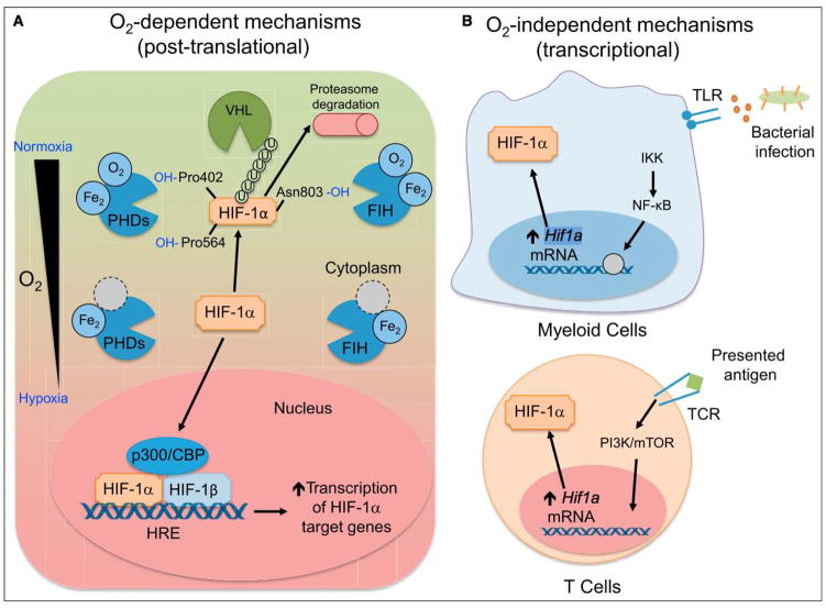
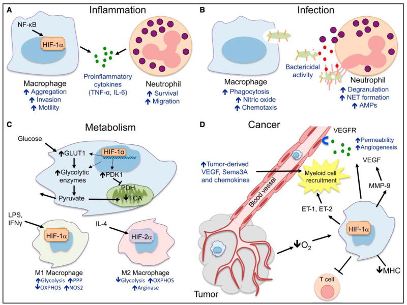
The hypoxic response in cells and tissues is mediated by the family of hypoxia-inducible factor (HIF) transcription factors; these play an integral role in the metabolic changes that drive cellular adaptation to low oxygen availability. HIF expression and stabilization in immune cells can be triggered by hypoxia, but also by other factors associated with pathological stress: e.g., inflammation, infectious microorganisms, and cancer. HIF induces a number of aspects of host immune function, from boosting phagocyte microbicidal capacity to driving T cell differentiation and cytotoxic activity. Cellular metabolism is emerging as a key regulator of immunity, and it constitutes another layer of fine-tuned immune control by HIF that can dictate myeloid cell and lymphocyte development, fate, and function. Here we discuss how oxygen sensing in the immune microenvironment shapes immunological response and examine how HIF and the hypoxia pathway control innate and adaptive immunity.


Figures

Figure 1
Figure 1. Mechanisms of HIF Stabilization by Immune Cells
(A) O2 dependent. When oxygen is available, HIF-1α is hydroxylated by PHD, enzymes that depend on oxygen and iron as cofactors. When prolylhydroxylated, HIF-1a is polyubiquitinated by VHL, marking it for proteasomal degradation. FIH hydroxylates HIF-1α at asparagine 803, which does not lead to polyubiquitination, but instead blocks interactions between HIF-1α and p300/CBP, a member of the HIF complex that acts as a coactivator of target gene transcription. When oxygen tension drops, PHD and FIH activity is inhibited, leading to HIF-1α accumulation and nuclear translocation, heterodimerization with HIF-1β, and recruitment of p300/CBP. The HIF transcriptional complex binds to hypoxia-response elements (HREs) to control target gene expression. (B) O2 independent. Bacterial products are recognized by TLRs expressed on myeloid cells, signaling through NF-κB to increase Hif1a transcription. In a similar fashion, TCR ligation upon antigen presentation on T lymphocytes leads to increased Hif1a transcription and HIF-1a protein accumulation, even in the presence of oxygen. While the mechanism of Hif1a mRNA induction is not known, activation of PI3K and mTOR appear to be involved in TCR-mediated induction of HIF-1α.
Figure 2
Figure 2. Hypoxia Pathway and Innate Immunity
(A) Inflammation. HIF-1α and NF-κB crosstalk regulates essential inflammatory functions in myeloid cells. HIF-1α increases macrophage aggregation, invasion, and motility and drives the expression of proinflammatory cytokines. HIF-1α also increases neutrophil survival by inhibiting apoptosis and triggers NF-κB-dependent neutrophilic inflammation. (B) Infection. HIF-1α increases intracellular bacterial killing by macrophages and also promotes granule protease production and release of nitric oxide (NO) and TNF-α, which in turn further contribute in antimicrobial control. (C) Cancer. As a result of hypoxia, HIF-1α is stabilized in cancer cells, increasing the production of chemotactic factors like CCL5, CXCL12, VEGF, endothelins ET-1 and ET-2, and semaphorin3A (Sema3A), which result in myeloid cell recruitment to the tumor. Once located in 31 hypoxic regions, tumor-associated macrophages stabilize HIF-1α, which contributes to tumor-promoting inflammation, angiogenesis, and impaired lymphocyte function. (D) Metabolism. HIF-1α in myeloid cells increases the transcription of key glycolytic enzymes, resulting on increased glucose uptake and glycolytic rate. Importantly, pyruvate entry in the citric acid cycle (TCA) is inhibited and converted to lactate, which is released to the extracellular compartment. This metabolic adaptation results in a decreased O2 consumption by limiting the rate of oxidative phosphorylation (OXPHOS). M1 and M2 polarized macrophages show different metabolic pathway preferences: whereas M1 rely on glycolysis as energy source, M2 have steady OXPHOS metabolism.
Figure 3
Figure 3. Hypoxia Pathway and Adaptive Immunity
(A) T cell differentiation. Upon activation, HIF-1α is strongly induced in conditions that favor differentiation of CD4+ Th17 cells. HIF-1α can increase the expression of RORgt, which in turn promotes IL-17 production and Th17 cell development. Th17 and Treg cells also rely on different metabolic pathways. HIF-1α can bind FOXP3 protein and impact its function and degradation, and in some cases, promotes Foxp3 mRNA induction and supports Treg cell function. HIF-1α-driven glycolytic shift supports Th17 cell differentiation, while lack of HIF-1α can support Treg cell differentiation under Th17 cell differentiation conditions. (B) HIF-1α stabilization by activated CD8+ T cells results in increased expression and release of important cytolytic molecules (granzyme B, perforin), increased expression of costimulatory/inhibitory molecules (CTLA-4, GITR, 4-1BB), and altered migration and chemokine receptor expression.

References

    1. Ohn GO, Seita J, Hong BJ, Kim YE, Bok S, Lee CJ, Kim KS, Lee JC, Leeper NJ, Cooke JP, et al. Transcriptional activation of hypoxia-inducible factor-1 (HIF-1) in myeloid cells promotes angiogenesis through VEGF and S100A8. Proc Natl Acad Sci USA. 2014;111:2698–2703. - PMC - PubMed
    1. Albina JE, Mastrofrancesco B, Vessella JA, Louis CA, Henry WL, Jr, Reichner JS. HIF-1 expression in healing wounds: HIF-1alpha induction in primary inflammatory cells by TNF-alpha. Am J Physiol Cell Physiol. 2001;281:C1971–C1977. - PubMed
    1. Anand RJ, Gribar SC, Li J, Kohler JW, Branca MF, Dubowski T, Sodhi CP, Hackam DJ. Hypoxia causes an increase in phagocytosis by macrophages in a HIF-1alpha-dependent manner. J Leukoc Biol. 2007;82:1257–1265. - PubMed
    1. Arany Z, Huang LE, Eckner R, Bhattacharya S, Jiang C, Goldberg MA, Bunn HF, Livingston DM. An essential role for p300/CBP in the cellular response to hypoxia. Proc Natl Acad Sci USA. 1996;93:12969–12973. - PMC - PubMed
    1. Baginska J, Viry E, Berchem G, Poli A, Noman MZ, van Moer K, Medves S, Zimmer J, Oudin A, Niclou SP, et al. Granzyme B degradation by autophagy decreases tumor cell susceptibility to natural killer-mediated lysis under hypoxia. Proc Natl Acad Sci USA. 2013;110:17450–17455. - PMC - PubMed

Publication types

Substances