. 2014 Nov 3:11:185.
doi: 10.1186/1743-422X-11-185.

Transmission of H7N9 influenza virus in mice by different infective routes

Affiliations

Transmission of H7N9 influenza virus in mice by different infective routes

Linlin Bao et al. Virol J. .

Erratum in

Abstract

Background: On 19 February 2013, the first patient infected with a novel influenza A H7N9 virus from an avian source showed symptoms of sickness. More than 349 laboratory-confirmed cases and 109 deaths have been reported in mainland China since then. Laboratory-confirmed, human-to-human H7N9 virus transmission has not been documented between individuals having close contact; however, this transmission route could not be excluded for three families. To control the spread of the avian influenza H7N9 virus, we must better understand its pathogenesis, transmissibility, and transmission routes in mammals. Studies have shown that this particular virus is transmitted by aerosols among ferrets.

Methods: To study potential transmission routes in animals with direct or close contact to other animals, we investigated these factors in a murine model.

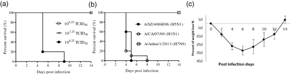
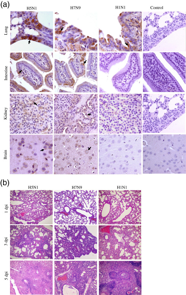
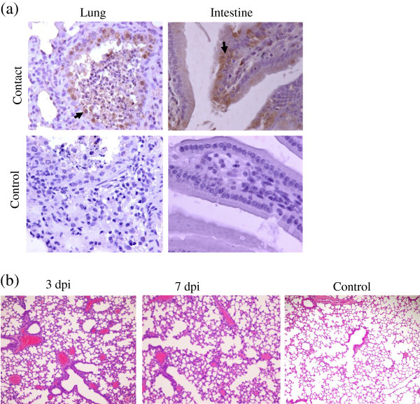
Results: Viable H7N9 avian influenza virus was detected in the upper and lower respiratory tracts, intestine, and brain of model mice. The virus was transmissible between mice in close contact, with a higher concentration of virus found in pharyngeal and ocular secretions, and feces. All these biological materials were contagious for naïve mice.

Conclusions: Our results suggest that the possible transmission routes for the H7N9 influenza virus were through mucosal secretions and feces.


Figures

Figure 1
Figure 1
Avian influenza virus H7N9 (A/Anhui/1/2013) infects mice. (a) LD50 of A/Anhui/1/2013 (H7N9). Representative results of three doses (108.25–106.25 TCID50) are shown. (b) Survival comparison of mice infected with H5N1, H1N1, or H7N9 viruses. (c) Percentage initial weight loss in mice infected with 106 TCID50 of H7N9 (A/Anhui/1/2013). Data are presented as the average values from two independent experiments ± SD (n =10 per group).
Figure 2
Figure 2
Histopathology and immunohistochemistry (IHC) analysis of infected mice. (a) Distribution of H7N9, H5N1, and H1N1 viruses in the tissues of infected mice as determined by IHC. Representative viral antigen distribution in tissues at 3 dpi is shown. Viral antigens are denoted with solid arrows (400× magnification). (b) Hematoxylin and eosin stain (HE) staining of lung tissues from infected mice (100× magnification).
Figure 3
Figure 3
H7N9 virus is transmissible among mice by direct contact. Seven naïve mice were placed in direct contact with three mice infected with 106 TCID50 of H7N9 virus. (a) IHC observations in tissues of mice exposed to H7N9-infected mice. Viral antigen distribution in tissues at 5 days following exposure to infected mice. Viral antigens are denoted with solid arrows (400× magnification). Viral antigens are within the epithelial cells of the bronchi and small intestinal villi, and within infiltrating inflammatory cells of the small intestine. (b) HE staining of lung tissue from mice that came in contact with H7N9-infected mice (100× magnification). At 3 days after coming into contact with infected mice, mild dilation of the interstitial pulmonary vasculature was observed in the lungs; congestion was observed after 7 days.
Figure 4
Figure 4
Transmission of influenza H7N9 virus among mice. Naïve mice were inoculated with throat secretions collected from infected mice. At 5 dpi, viral antigens were found in epithelial cells of the lungs, gastric and intestinal mucosa, and in infiltrating inflammatory cells of the small intestine (solid arrows; 400× magnification). At 5 dpi, limited interstitial pneumonia and inflammatory cell infiltration, degeneration and necrosis of epithelial cells in the gastric mucosa, and vacuolar degeneration of epithelial cells in small intestine villi was seen (100× magnification).

Similar articles

Cited by

References

    1. Gao R, Cao B, Hu Y, Feng Z, Wang D, Hu W, Chen J, Jie Z, Qiu H, Xu K, Xu X, Lu H, Zhu W, Gao Z, Xiang N, Shen Y, He Z, Gu Y, Zhang Z, Yang Y, Zhao X, Zhou L, Li X, Zou S, Zhang Y, Li X, Yang L, Guo J, Dong J, Li Q, et al. Human Infection with a Novel Avian-Origin Influenza A (H7N9) Virus. N Engl J Med. 2013;368(20):1888–1897. doi: 10.1056/NEJMoa1304459. - DOI - PubMed
    1. Horby P. H7N9 is a virus worth worrying about. Nature. 2013;496:399. doi: 10.1038/496399a. - DOI - PubMed
    1. Koopmans M, de Jong MD. Avian influenza A H7N9 in Zhejiang, China. Lancet. 2013;381(9881):1882–1883. doi: 10.1016/S0140-6736(13)60936-8. - DOI - PubMed
    1. Wu S, Wu F, He J. Emerging risk of H7N9 influenza in China. Lancet. 2013;381:1539–1540. doi: 10.1016/S0140-6736(13)60767-9. - DOI - PMC - PubMed
    1. Zhang W, Wang L, Hu W, Ding F, Sun H, Li S, Huang L, Li C. Epidemiological characteristics of cases for influenza A (H7N9) virus infections in China. Clin Infect Dis. 2013;57(4):619–620. doi: 10.1093/cid/cit277. - DOI - PMC - PubMed

Publication types