. 2014 Nov 3;2(11):e12185.
doi: 10.14814/phy2.12185. Print 2014 Nov 1.

Stand-up exercise training facilitates muscle recovery from disuse atrophy by stimulating myogenic satellite cell proliferation in mice

Affiliations

Stand-up exercise training facilitates muscle recovery from disuse atrophy by stimulating myogenic satellite cell proliferation in mice

Yuta Itoh et al. Physiol Rep. .

Abstract

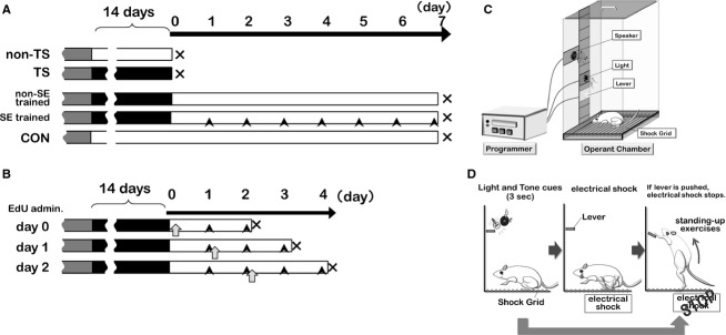
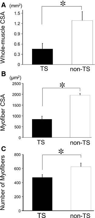
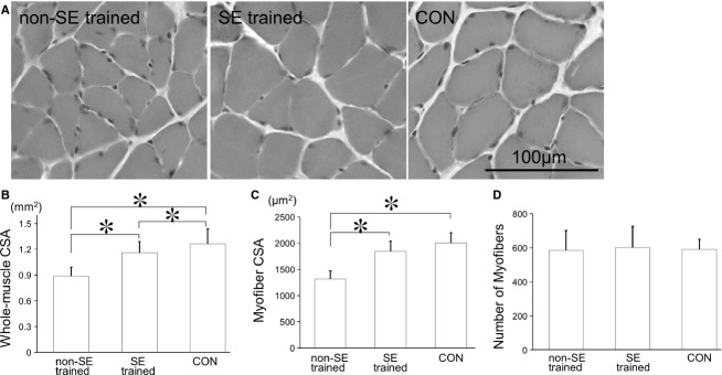
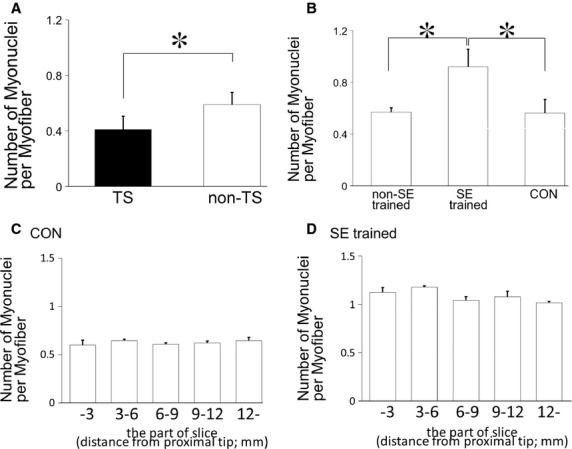
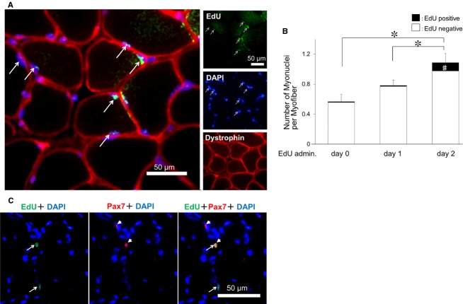
Determining the cellular and molecular recovery processes in inactivity - or unloading -induced atrophied muscles should improve rehabilitation strategies. We assessed the effects of stand-up exercise (SE) training on the recovery of atrophied skeletal muscles in male mice. Mice were trained to stand up and press an elevated lever in response to a light-tone cue preceding an electric foot shock and then subjected to tail suspension (TS) for 2 weeks to induce disuse atrophy in hind limb muscles. After release from TS, mice were divided into SE-trained (SE cues: 25 times per set, two sets per day) and non-SE-trained groups. Seven days after the training, average myofiber cross-sectional area (CSA) of the soleus muscle was significantly greater in the SE-trained group than in the non-SE-trained group (1843 ± 194 μm(2) vs. 1315 ± 153 μm(2)). Mean soleus muscle CSA in the SE trained group was not different from that in the CON group subjected to neither TS nor SE training (2005 ± 196 μm(2)), indicating that SE training caused nearly complete recovery from muscle atrophy. The number of myonuclei per myofiber was increased by ~60% in the SE-trained group compared with the non-SE-trained and CON groups (0.92 ± 0.03 vs. 0.57 ± 0.03 and 0.56 ± 0.11, respectively). The number of proliferating myonuclei, identified by 5-ethynyl-2'-deoxyuridine staining, increased within the first few days of SE training. Thus, it is highly likely that myogenic satellite cells proliferated rapidly in atrophied muscles in response to SE training and fused with existing myofibers to reestablish muscle mass.

Keywords: 5‐ethynyl‐2’‐deoxyuridine; myogenic satellite cells; myonuclei; operant conditioning; stand‐up exercise.


Figures

Figure 1.
Figure 1.
Scheme for intervention times and experimental methods. (A) Treatment protocols for mouse groups: gray, operant conditioning; black, tail suspension (TS); open, normal housing; black arrowhead, stand‐up exercise (SE) training. (B) During SE training, mice were administered 5‐ethynyl‐2’‐deoxyuridine on day 0, 1, or 2 of the SE‐training period (open arrows), and sacrificed 48 h later. (C) Diagram of the operant‐conditioning device. (D) Learning program for stand‐up training. An electrical shock was generated in a shock grid at 3 sec after displaying light and tone cues. The electrical shock was stopped when a mouse pushed the lever. The electrical shock was not used when a mouse pushed the lever in response to the cues before a shock was generated. The mice acquired the stand‐up exercise without an electrical shock after 7 days of learning (100 times/day).
Figure 2.
Figure 2.
Changes in muscle histology after prolonged tail suspension (TS). (A) Whole‐muscle cross‐sectional area (CSA), (B) myofiber CSA, and (C) the number of myofibers after hind limb unloading using prolonged TS. Whole‐muscle CSA, myofiber CSA, and the number of myofibers were significantly reduced in the TS group compared with the control non‐TS group. Solid bars, TS group; open bars, non‐TS group. Results are means ± SEM's; n = 6 per group. *P <0.05.
Figure 3.
Figure 3.
Changes in muscle histology after stand‐up exercise (SE) training. (A) Photomicrographs of muscle samples (hematoxylin–eosin stained) from mice subjected to TS but not SE training (non‐SE‐trained group), subjected to TS and then SE training (SE‐trained group), and control mice (CON). (B) Whole‐muscle CSA, (C) myofiber CSA, and (D) the number of myofibers for these three groups. Myofiber CSA was significantly greater in the SE‐trained group than in the non‐SE‐trained group and approximately the same as in the CON group. There were no significant differences in the numbers of myofibers among these three groups. Results are means ± SEM's; n = 6 per group. *P <0.05.
Figure 4.
Figure 4.
SE‐training effects on numbers of myonuclei in atrophied muscles. (A, B) Numbers of myonuclei per myofiber after TS (A) and SE training (B). There were significantly fewer myonuclei per myofiber in the TS group compared with the control non‐TS group, and there were significantly more myonuclei per myofiber in the SE‐trained group compared with the non‐SE‐trained and CON groups. Results are means ± SEM's; n = 6 per group. *P <0.05. (C, D) The numbers of myonuclei per myofiber were counted in different cross sections of soleus muscles: proximal tip–3 mm, 3–6 mm, 6–9 mm, 9–12 mm, and 12 mm–distal tip. Between these different sections, there were no significant differences in the numbers of myonuclei in the CON (B) and SE trained (C) groups. Results are means ± SEM's; n = 3 for each muscle part.
Figure 5.
Figure 5.
SE‐training‐induced increases in myonuclei are due to the proliferation and fusion of myogenic satellite cells. (A) Typical section stained by 5‐ethynyl‐2’‐deoxyuridine (EdU), antidystrophin, and 4’,6‐diamidino‐2‐phenylindole. (B) Numbers of myonuclei per myofiber and EdU‐positive myonuclei (A, arrow). The number of myonuclei per myofiber was highest in the group administered EdU on day 2 of SE training. *P <0.05 for total myonuclei counts, #P <0.05 for EdU‐positive myonuclei counts. (C) Photomicrographs of muscle samples stained with an anti‐Pax7 antibody (red, arrowheads), a marker of myogenic satellite cells, from mice subjected to TS and then SE training (SE‐trained group). Immunoreactivity to Pax7 was observed in EdU‐positive nuclei (green, arrows).

Similar articles

Cited by

References

    1. Antonio J., Gonyea W. J. 1993a. Skeletal muscle fiber hyperplasia. Med. Sci. Sports Exerc.; 25:1333-1345. - PubMed
    1. Antonio J., Gonyea W. J. 1993b. Progressive stretch overload of skeletal muscle results in hypertrophy before hyperplasia. J. Appl. Physiol.; 75:1263-1271. - PubMed
    1. Bischoff R. 1989. Analysis of muscle regeneration using single myofibers in culture. Med. Sci. Sports Exerc.; 215 Suppl:S164-S172. - PubMed
    1. Bruusgaard J. C., Johansen I. B., Egner I. M., Rana Z. A., Gundersen K. 2010. Myonuclei acquired by overload exercise precede hypertrophy and are not lost on detraining. Proc. Natl Acad. Sci. USA; 107:15111-15116. - PMC - PubMed
    1. Bruusgaard J. C., Egner I. M., Larsen T. K., Dupre‐Aucouturier S., Desplanches D., Gundersen K. 2012. No change in myonuclear number during muscle unloading and reloading. J. Appl. Physiol.; 113:290-296. - PubMed

LinkOut - more resources