Review
. 2014 Nov 3;5(2):a019679.
doi: 10.1101/cshperspect.a019679.

Fungal morphogenesis

Affiliations
Review

Fungal morphogenesis

Xiaorong Lin et al. Cold Spring Harb Perspect Med. .

Abstract

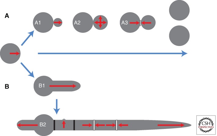
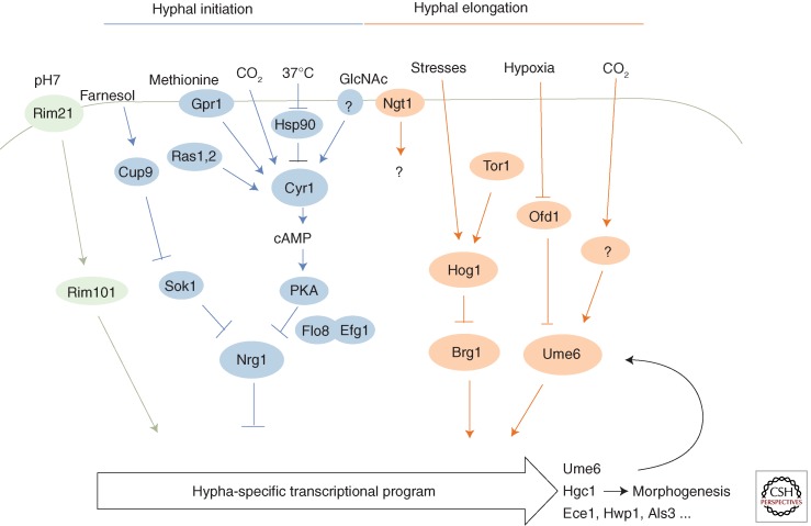
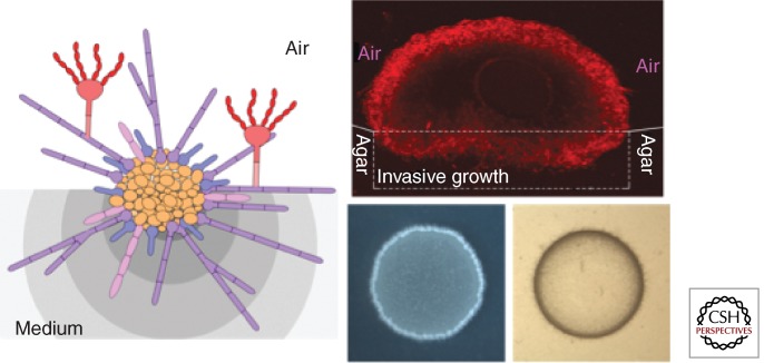
Morphogenesis in fungi is often induced by extracellular factors and executed by fungal genetic factors. Cell surface changes and alterations of the microenvironment often accompany morphogenetic changes in fungi. In this review, we will first discuss the general traits of yeast and hyphal morphotypes and how morphogenesis affects development and adaptation by fungi to their native niches, including host niches. Then we will focus on the molecular machinery responsible for the two most fundamental growth forms, yeast and hyphae. Last, we will describe how fungi incorporate exogenous environmental and host signals together with genetic factors to determine their morphotype and how morphogenesis, in turn, shapes the fungal microenvironment.


Figures

Figure 1.
Figure 1.
Distinct patterns of morphogenesis in yeast and hyphal cells. Yeasts and filamentous fungi typically initiate growth as nonpolarized cells or spores. Budding yeasts such as Saccharomyces cerevisiae or Cryptococcus neoformans (A) establish an axis of polarity that directs the emergence of a new bud (A1). Following a period of polarized growth, depolarization enables the formation of an ellipsoidal bud (A2). Following nuclear division, the construction and controlled degradation of a septum (A3) results in cell separation. Filamentous fungi such Aspergillus nidulans and Candida albicans (B) establish a polarity axis that directs the emergence of a germ tube (B1). Unlike yeasts, sustained polar growth leads to the formation of a hypha that grows by apical extension. Moreover, hyphae are able to simultaneously support multiple polarity axes to allow the formation of septal cross-walls (bars) and lateral branches (B2). For many filamentous fungi, spores also generate secondary germ tubes once they are partitioned from the primary hypha by a septum.
Figure 2.
Figure 2.
C. neoformans Rac2 localization suggests a role in cell polarity. A Gfp–Rac2 fusion protein was expressed in C. neoformans and visualized using an Olympus (Center Valley, PA) IX70 microscope. After image acquisition and deconvolution of serial images in the Z-coordinate using Deltavision (GE Healthcare, Issaquah, WA) software, pseudocolored, merged images were produced using Fiji (Madison, WI) software. The most intense fluorescent signal, representing enrichment of Rac2 localization, is present at the site of incipient bud emergence (arrow) and distal edge of a small daughter cell (arrowhead). (Image provided by S. Esher and K. Selvig, Duke University.)
Figure 3.
Figure 3.
Signal transduction pathways integrating various signals for morphogenesis in C. albicans. Selective signal transduction pathways and regulators of morphogenesis are shown. Arrows indicate activation. Bars indicate inhibition. Hyphal-specific regulator Ume6 sustains the hypha-specific transcriptional program and Hgc1 promotes hyphal morphogenesis.
Figure 4.
Figure 4.
A fungal colony is a heterogeneous population. The left panel shows a diagram of a fungal colony composed of multiple morphotypes based on what is known about Cryptococcus. Yeast cells (orange) populate the colony center, mixed with some pseudohyphae (pink), germ tubes (blue), and hyphae (purple), which predominate at the periphery of the colony. The filamentous cells also invade the medium (invasive hyphae) or extend into the air (aerial hyphae). Some aerial hyphae further develop into fruiting bodies and produce sexual spores (red). Different microenvironments in the medium are indicated with different shades of gray. Different cell types have different cell surface composition and structure. Even for the same morphotype, cells are phenotypically and physiologically different, based on their microenvironment and spatial position. The right upper panel shows a confocal image of a Cryptococcus colony derived from a single yeast cell. The bifunctional adhesion protein Cfl1 fused with m-Cherry fluorescent protein is highly expressed in the hyphal subpopulation located at the colony periphery. Yeast cells appear dark because of low levels of Cfl1 expression. The right lower panel shows the colony that developed after a yeast-cell suspension was dropped on the filamentation agar. The white fluffy appearance at the colony edge is caused by the presence of aerial hyphae. Invasive growth, detected as cells that remain when the surface is washed, is shown on the bottom right. The invasive cells, mostly in the germ tube or hyphal form at the periphery of the colony, penetrate into agar medium forming a ring-like footprint. (Images provided by L. Wang and X. Tian, Texas A&M University.)

References

    1. Adams DJ 2004. Fungal cell wall chitinases and glucanases. Microbiol 150: 2029–2035. - PubMed
    1. Aimanianda V, Bayry J, Bozza S, Kniemeyer O, Perruccio K, Elluru SR, Clavaud C, Paris S, Brakhage AA, Kaveri SV, et al. 2009. Surface hydrophobin prevents immune recognition of airborne fungal spores. Nature 460: 1117–1121. - PubMed
    1. Almeida RS, Brunke S, Albrecht A, Thewes S, Laue M, Edwards JE, Filler SG, Hube B 2008. The hyphal-associated adhesin and invasin Als3 of Candida albicans mediates iron acquisition from host ferritin. PLoS Pathog 4: e1000217. - PMC - PubMed
    1. Alonso-Monge R, Navarro-Garcia F, Molero G, Diez-Orejas R, Gustin M, Pla J, Sanchez M, Nombela C 1999. Role of the mitogen-activated protein kinase Hog1p in morphogenesis and virulence of Candida albicans. J Bacteriol 181: 3058–3068. - PMC - PubMed
    1. Alonso-Monge R, Roman E, Arana DM, Prieto D, Urrialde V, Nombela C, Pla J 2010. The Sko1 protein represses the yeast-to-hypha transition and regulates the oxidative stress response in Candida albicans. Fungal Genet Biol 47: 587–601. - PubMed