Review
. 2014 Nov 3;4(11):a013912.
doi: 10.1101/cshperspect.a013912.

How to make a heart valve: from embryonic development to bioengineering of living valve substitutes

Affiliations
Review

How to make a heart valve: from embryonic development to bioengineering of living valve substitutes

Donal MacGrogan et al. Cold Spring Harb Perspect Med. .

Abstract

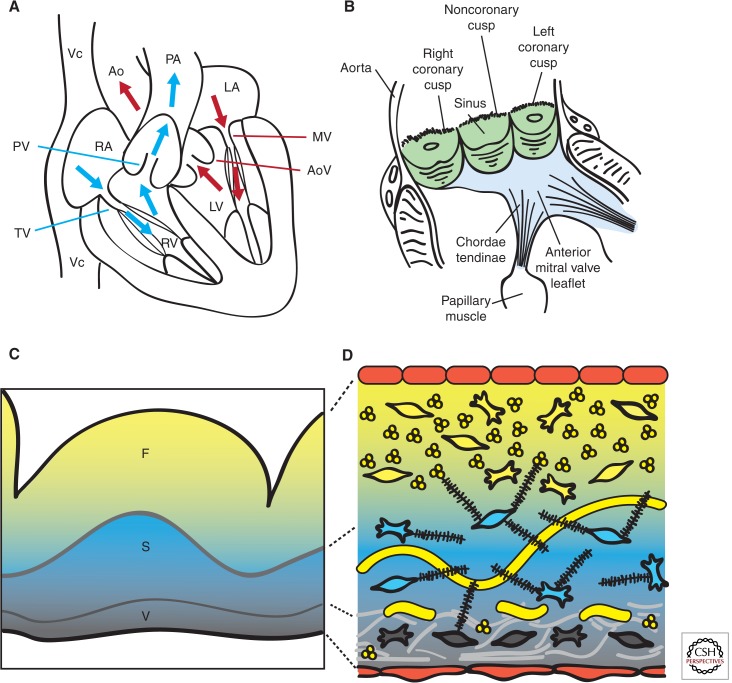
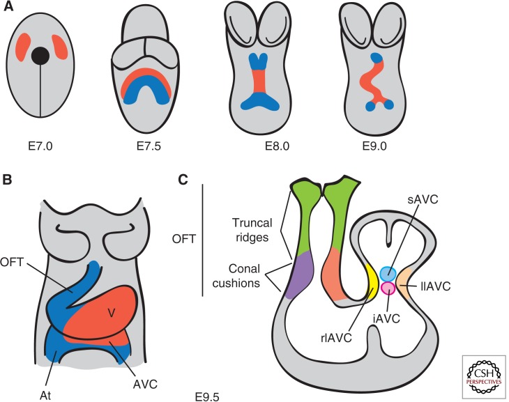
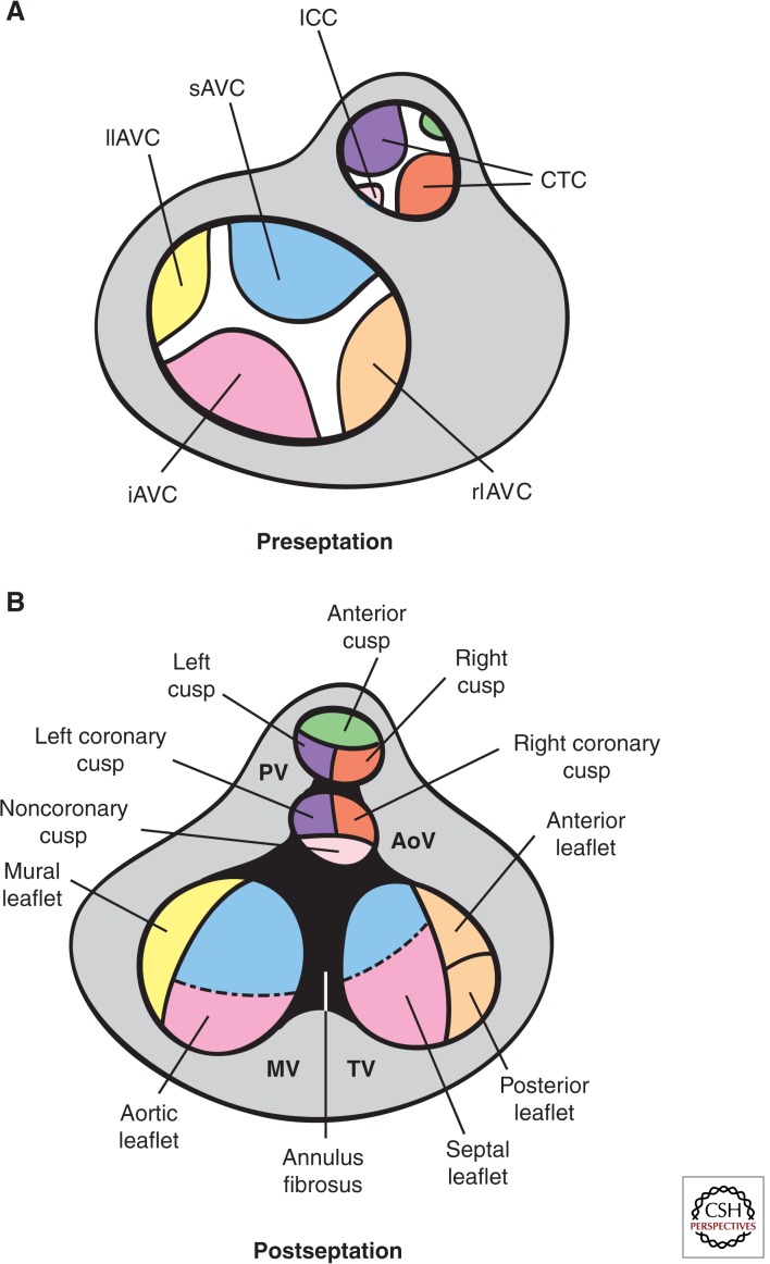
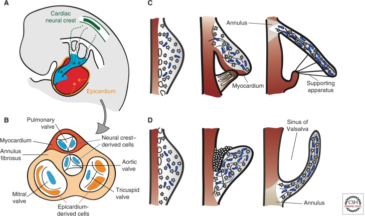
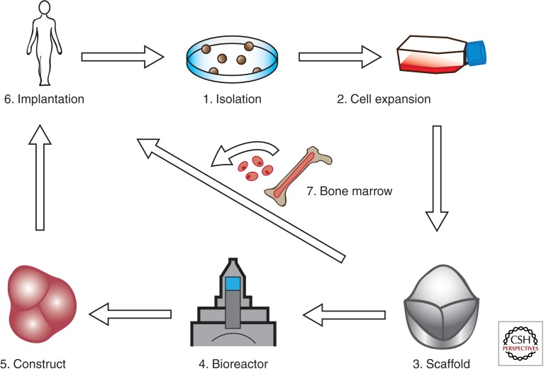
Cardiac valve disease is a significant cause of ill health and death worldwide, and valve replacement remains one of the most common cardiac interventions in high-income economies. Despite major advances in surgical treatment, long-term therapy remains inadequate because none of the current valve substitutes have the potential for remodeling, regeneration, and growth of native structures. Valve development is coordinated by a complex interplay of signaling pathways and environmental cues that cause disease when perturbed. Cardiac valves develop from endocardial cushions that become populated by valve precursor mesenchyme formed by an epithelial-mesenchymal transition (EMT). The mesenchymal precursors, subsequently, undergo directed growth, characterized by cellular compartmentalization and layering of a structured extracellular matrix (ECM). Knowledge gained from research into the development of cardiac valves is driving exploration into valve biomechanics and tissue engineering directed at creating novel valve substitutes endowed with native form and function.


Figures

Figure 1.
Figure 1.
Heart valve structure and function. (A) Oxygen-depleted blood (blue) enters the heart from the vena cava (Vc) through the right atrium (RA) and transits via the tricuspid valve (TV) into the right ventricle (RV). From there, it passes through the pulmonary valve (PV) and reaches the lungs via the pulmonary artery (PA). Oxygen-rich blood from the lungs (red) enters the left atrium (LA) and empties into the left ventricle (LV) by passing through the mitral valve (MV). From the LV, the blood is pumped through the aortic valve (AoV) to the aorta (Ao), and from there it distributes to the rest of the body. (B) Scheme of a longitudinally opened aortic root, showing the close relation between aortic (green) and mitral (blue) valves in situ. The cusps are attached to the annulus fibrosus, a fibroelastic structure entrapping the valves (not shown). The free edge of the AoV leaflets is anchored to the LV wall by papillary muscles via chordae tendinae. (C) Artist rendition of a Movat’s pentachrome staining of a cross section of the AoV cusp. (D) Depiction of the detailed architecture. The monolayer of endothelium (red) lining the valves has a cuboidal appearance on the arterial side and a flattened appearance of the ventricular side. Fibroblasts, smooth muscle, and myofibrolasts are interspersed in the different layers. Collagen (yellow) is densely packed and fibrillar and circumferential in the fibrosa (F), loose in the spongiosa (S), and multidirectional in the ventricularis (V). Proteoglycans (black) predominate in the spongiosa, and interact with cells and collagen. Radial elastin (light gray) predominates in the ventricularis.
Figure 2.
Figure 2.
Early stages of heart development. (A) Ventral views of the developing mouse embryo. At E7.0, cardiac progenitors (red) have reached the head folds and, by E7.5, two cardiac lineages can be distinguished: the first heart field (FHF) (red) and the second heart field (SHF) (blue). At E8.0, the FHF progenitors merge to form the heart tube, which elongates at arterial and venous poles by the addition of progenitor cells from the second heart field (SHF). Between E8.0 and E9.0, the heart tube undergoes rightward looping. (B) Ventral view of the E9.5 heart, which consists of four anatomically distinct regions: atrium (At), atrioventricular canal (AVC), ventricle (V), and the outflow tract (OFT). (C) Longitudinal section depicting the prevalvular ECs. Two elongated cushions can be seen in the OFT, consisting of proximal (conal cushions, purple) and distal (truncal ridges, green) sections. The AVC has four cushions: right lateral (rlAVC), left lateral (llAVC), superior (sAVC), and inferior (iAVC). (Figure created from data adapted from Snarr et al. 2008.)
Figure 3.
Figure 3.
Fate of the cardiac cushions in the mature heart. Top view of the OFT preseptation (A) and postseptation (B). In the AVC, fusion of the major and lateral AV cushions at E13.5 results in formation of inlet valves. The sAVC (blue) contributes mostly to the aortic leaflet of the MV, whereas the iAVC (pink) contributes mostly to the septal leaflet of the TV. The rlAVC (orange) and llAVC (yellow) contribute, respectively, to the mural leaflet of the MV and anterior and posterior leaflets of the TV. These leaflets are later invaded by epicardially derived mesenchyme. The conotruncal cushions (CTCs) develop laterally right (ocre) and left (purple) in the OFT and are separated by smaller anterior (green) and posterior (light pink) intercalated cushions (ICCs). At E12.5, fusion of the CTCs yields the mesenchymal outlet septum, the right and left (R–L) cusps of the PV and R–L coronary cusps of the AoV. The ICCs give rise to the anterior leaflet of the PV and the noncoronary cusps of the AoV. Neural crest precursors invading the distal truncal ridges participate in OFT septation and contribute mesenchyme to the R–L cusps of both PV and AV. (Figure created from data adapted from Snarr et al. 2008 and Lin et al. 2012.)
Figure 4.
Figure 4.
Remodeling of the cardiac valve leaflets and cusps. (A) Contribution of cardiac neural crest (green)- and epicardial (orange)- derived cells (CNCC and EPDCs). CNCCs migrate through the pharyngeal arches and into the OFT to initiate the reorganization of the OFT and formation of SL valves. EPDCs contribute to the formation of the coronary arteries, interstitial cells in the myocardium, and the AV valves. (B) Top view of the septated heart, depicting the relative contributions of CNCCs and EPDCs to cardiac valve leaflets and cusps. (C) Morphogenesis of the AV valves. From E13.5 onward, the leaflets and tensile apparatus (light brown) of the AV valves form predominantly by delamination of the inner layers of the inlet zone from the ventricular septal wall. The chordae tendinae, annulus fibrosus, and the leaflet itself are derived from the endocardium. The septal leaflet of the TV (in contrast to the aortic leaflet of the MV), together with its supporting tendinous cords, remains connected to the myocardium until E17.5. (D) Morphogenesis of the SL valves. The excavation of the cusps takes place initially by solid ingrowth of the endothelium at the arterial face of the cusp and, subsequently, by lumenation. Elongation and remodeling of the primordia into mature valve structures are associated with regionalized cell proliferation and matrix alignment guided by hemodynamic forces. Myocardium is depicted in dark red, proteglycans in blue, and mesenchyme in gray.
Figure 5.
Figure 5.
Valve tissue–engineering paradigm(s). (1)–(3) Conventional valve bioengineering entails a scaffold that is seeded with cells to grow a valve construct ex vivo in a bioreactor (dynamic conditioning) (4), followed by implantation in the anatomic site in vivo (5). In the modified protocol, the appropriate scaffold (3) is implanted in vivo (6) and cellularization is achieved by recruitment of circulating endothelial and mesenchymal cells from bone marrow (7). The scaffold can be engineered to incorporate molecules that stimulate recruitment, adhesion, migration, proliferation, differentiation, and cell function within the scaffold. (Figure created from data adapted from Weber et al. 2012.)

Similar articles

Cited by

References

    1. Abu-Issa R, Kirby ML. 2007. Heart field: From mesoderm to heart tube. Annu Rev Cell Dev Biol 23: 45–68 - PubMed
    1. Aikawa E, Whittaker P, Farber M, Mendelson K, Padera RF, Aikawa M, Schoen FJ. 2006. Human semilunar cardiac valve remodeling by activated cells from fetus to adult: Implications for postnatal adaptation, pathology, and tissue engineering. Circulation 113: 1344–1352 - PubMed
    1. Akiyama H, Chaboissier MC, Behringer RR, Rowitch DH, Schedl A, Epstein JA, de Crombrugghe B. 2004. Essential role of Sox9 in the pathway that controls formation of cardiac valves and septa. Proc Natl Acad Sci 101: 6502–6507 - PMC - PubMed
    1. Alfieri CM, Cheek J, Chakraborty S, Yutzey KE. 2010. Wnt signaling in heart valve development and osteogenic gene induction. Dev Biol 338: 127–135 - PMC - PubMed
    1. Anderson RH, Webb S, Brown NA, Lamers W, Moorman A. 2003. Development of the heart: (3) Formation of the ventricular outflow tracts, arterial valves, and intrapericardial arterial trunks. Heart 89: 1110–1118 - PMC - PubMed

Publication types