. 2015 Apr;64(4):1420-30.
doi: 10.2337/db14-0872. Epub 2014 Nov 3.

Epigenetic changes in bone marrow progenitor cells influence the inflammatory phenotype and alter wound healing in type 2 diabetes

Affiliations

Epigenetic changes in bone marrow progenitor cells influence the inflammatory phenotype and alter wound healing in type 2 diabetes

Katherine A Gallagher et al. Diabetes. 2015 Apr.

Abstract

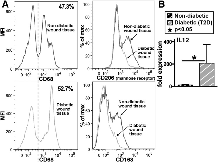
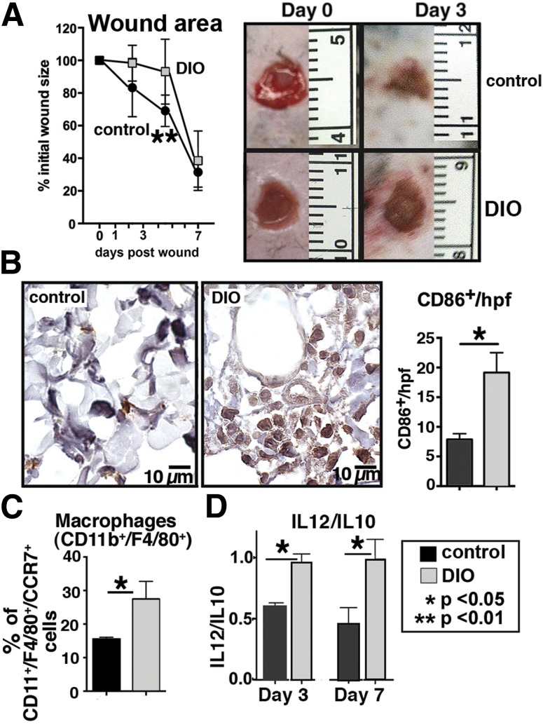
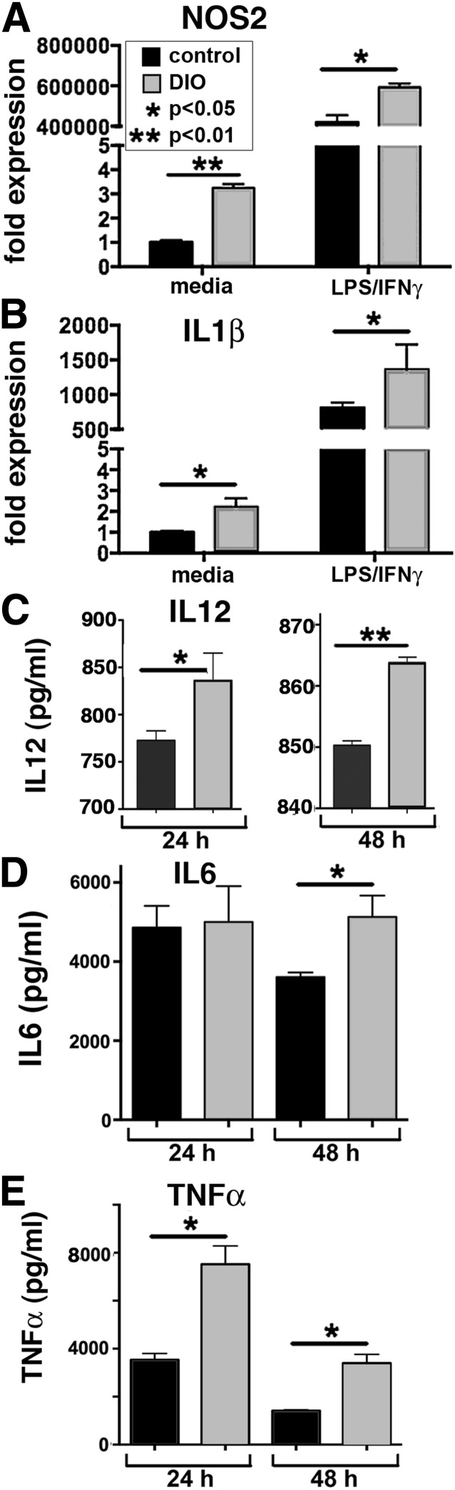
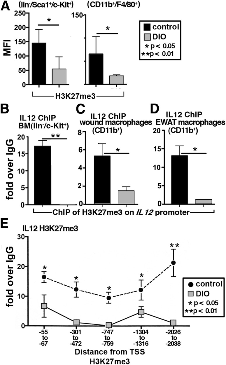
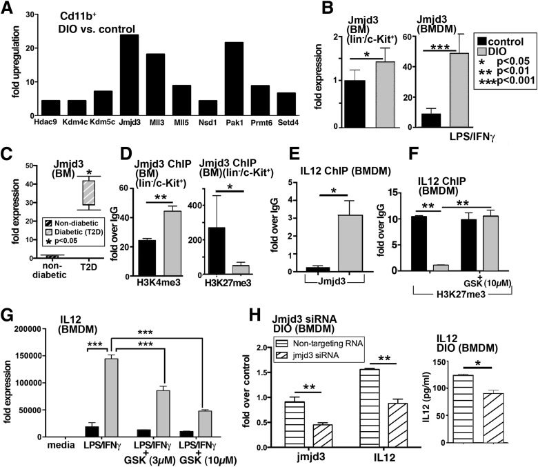
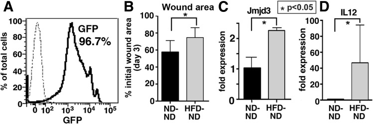
Classically activated (M1) macrophages are known to play a role in the development of chronic inflammation associated with impaired wound healing in type 2 diabetes (T2D); however, the mechanism responsible for the dominant proinflammatory (M1) macrophage phenotype in T2D wounds is unknown. Since epigenetic enzymes can direct macrophage phenotypes, we assessed the role of histone methylation in bone marrow (BM) stem/progenitor cells in the programming of macrophages toward a proinflammatory phenotype. We have found that a repressive histone methylation mark, H3K27me3, is decreased at the promoter of the IL-12 gene in BM progenitors and this epigenetic signature is passed down to wound macrophages in a murine model of glucose intolerance (diet-induced obese). These epigenetically "preprogrammed" macrophages result in poised macrophages in peripheral tissue and negatively impact wound repair. We found that in diabetic conditions the H3K27 demethylase Jmjd3 drives IL-12 production in macrophages and that IL-12 production can be modulated by inhibiting Jmjd3. Using human T2D tissue and murine models, we have identified a previously unrecognized mechanism by which macrophages are programmed toward a proinflammatory phenotype, establishing a pattern of unrestrained inflammation associated with nonhealing wounds. Hence, histone demethylase inhibitor-based therapy may represent a novel treatment option for diabetic wounds.


Figures

Figure 1
Figure 1
Human wound and BMDMs exhibit decreased anti-inflammatory (M2-like) and increased proinflammatory (M1-like) characteristics in T2D. A: Flow cytometry analysis of human macrophages (CD68+) isolated from nonischemic, nondiabetic, and T2D chronic wound tissue. Cells were stained for CD163 and mannose receptor (CD206). Percentage of CD68+ cells expressing either CD206 (top right) or CD163 (bottom right) are shown (n = 6). B: RT-PCR quantification of IL-12 (M1 cytokine) expression in human BMDMs 6 h following stimulation with LPS/IFN-γ. BMDMs from T2D patients are compared with BMDMs from nondiabetic patients (n = 6). Data are expressed as mean ± SE. MFI, mean fluorescence intensity.
Figure 2
Figure 2
Delayed wound healing in DIO mice is associated with increased proinflammatory macrophages. Punch biopsies (4 mm) were performed on the back of DIO and control mice. Change in wound area was recorded daily using ImageJ software (National Institutes of Health) until complete healing was observed. A: Wound healing curves in DIO and control mice (n = 10/time point). Data are pooled from three experiments and are expressed as mean ± SD. Representative images of wounds at day 0 and 3 days postwounding are shown. B: Immunohistochemical analysis of mononuclear cells (CD86+) in wounds at day 3 (cells/high-powered field [hpf]) in DIO compared with controls (n = 6). Representative examples of DIO and control wounds are shown. C: Flow cytometry of DIO and control wounds at day 3. Proinflammatory macrophages were defined as CD11b+/F4/80+ cells that coexpressed CCR7 (n = 6, experiment replicated once). D: Ratio of IL-12 (M1) to IL-10 (M2) cytokine levels in wounds at days 3 and 7 analyzed by Bioplex (n = 4, experiment replicated one time). Data are expressed as mean ± SE.
Figure 3
Figure 3
Macrophages are predisposed toward a proinflammatory (M1) phenotype in a T2D murine model. A and B: NOS2 and IL-1β expression levels quantified by RT-PCR in DIO and control BMDMs at 6 and 24 h after stimulation with LPS/IFN-γ (n = 4, replicated two times). C: Levels of IL-12 in supernatants from DIO and control BMDMs following treatment with LPS/IFN-γ at 24 and 48 h as measured by ELISA (n = 4, plated in triplicate). D and E: Levels of IL-6 and TNF-α in supernatants from DIO and control BMDMs following treatment with LPS/IFN-γ at 24 and 48 h as measured by Bioplex. Protein levels are expressed as pg/mL (n = 4, plated in triplicate). Data are expressed as mean ± SE.
Figure 4
Figure 4
DIO BM stem/progenitor cells and peripheral macrophages display decreased trimethylation of H3K27me3 on the IL-12 promoter. A: H3K27me3 levels in BM stem/progenitor cells (lin/Sca1+/c-Kit+) and BMDMs (CD11b+/F4/80+) from DIO and control mice measured by flow cytometry (n = 4). BD: BM stem/progenitor cells (lin/c-Kit+), wound, and EWAT macrophages (CD11b+) were isolated in vivo using MACS and analyzed by ChIP for H3K27me3 levels on the promoter of IL-12 (n = 3/group, plated in triplicate). E: H3K27me3 methylation was measured along the IL-12 promoter in BMDMs (n = 3, replicated twice). Data are expressed as mean ± SE. MFI, mean fluorescence intensity; TSS, transcription start site.
Figure 5
Figure 5
Increased JmjC demethylase, Jmjd3, production in BM LK cells and macrophages. A: Gene expression of chromatin-modifying enzymes was analyzed using PCR array plates in CD11b+ MACS-isolated cells from DIO and control wounds at day 3. Enzymes that were upregulated are shown. The threshold was set to a fourfold difference. Data are expressed as fold over control (n = 3/group). B: RT-PCR quantification of Jmjd3 levels in DIO and control BM progenitor cells (lin/c-Kit+) (isolated by MACS) and BMDMs (following stimulation with LPS/IFN-γ) (n = 4/group). C: RT-PCR analysis of human BM isolated from nondiabetic and T2D patients undergoing amputations at the femur level (n = 2/group). D: ChIP analysis of H3K4me3 and H3K27me3 on the Jmjd3 promoter of in vivo MACS-isolated BM LK cells from DIO and control mice (n = 5/group). E: ChIP analysis for Jmjd3 on the IL-12 promoter in DIO BMDMs compared with controls (n = 3/group, replicated once). F: BMDMs from DIO and control mice were treated with the H3K27me3 demethylase inhibitor, GSK-J4 (10 µmol/L), for 6 h, and ChIP analysis of H3K27me3 on the IL-12 promoter was performed (n = 3/group). G: BMDMs from DIO and control mice were stimulated with LPS/IFN-γ in the presence or absence of GSK-J4 (3 µmol/L, 10 μmol/L) for 6 h, and analysis of IL-12 transcription was performed (n = 3/group). Data are expressed as mean ± SE. H: DIO BMDMs were transfected with Jmjd3 siRNA or nontargeting siRNA or lipofectamine alone in triplicate wells. Forty-eight hours later, cells were stimulated with LPS/IFN-γ for 24 h. Transcript levels of Jmjd3 and IL-12 in cells treated with Jmjd3 siRNA or nontargeting siRNA are shown. Following stimulation, cell supernatants were collected and used for IL-12 Bioplex. Results are expressed as mean ± SE. Statistical analysis was performed using Student t tests, and P value <0.05 was considered significant.
Figure 6
Figure 6
Wound healing is impaired and macrophage function is altered in GFP+DIO BM chimeras. BM chimeras were created using GFP+ mice on a C57BL/6 background. GFP+ mice were fed an HFD (60% fat) or ND (12% fat) for 14 weeks, and BM from these mice was transferred into irradiated C57BL/6 recipients. A: Peripheral blood analysis was performed weekly and at 8 weeks confirmed 96.5% donor chimerism. B: Change in wound area compared with initial wound size at day 3 postwounding (n = 4/group). C and D: Jmjd3 and IL-12 expression in in vivo macrophages (CD11b+) MACS isolated from wounds at day 3 were quantified by RT-PCR in ND→ND and HFD→ND GFP+ chimeric mice (n = 4/group). Data are expressed as mean ± SE.

Comment in

References

    1. Driver VR, Fabbi M, Lavery LA, Gibbons G. The costs of diabetic foot: the economic case for the limb salvage team. J Vasc Surg 2010;52(Suppl.):17S–22S - PubMed
    1. Fathke C, Wilson L, Hutter J, et al. . Contribution of bone marrow-derived cells to skin: collagen deposition and wound repair. Stem Cells 2004;22:812–822 - PMC - PubMed
    1. Okuno Y, Nakamura-Ishizu A, Kishi K, Suda T, Kubota Y. Bone marrow-derived cells serve as proangiogenic macrophages but not endothelial cells in wound healing. Blood 2011;117:5264–5272 - PMC - PubMed
    1. Sica A, Mantovani A. Macrophage plasticity and polarization: in vivo veritas. J Clin Invest 2012;122:787–795 - PMC - PubMed
    1. Martin P. Wound healing—aiming for perfect skin regeneration. Science 1997;276:75–81 - PubMed

Publication types