. 2014 Nov 18;111(46):16604-9.
doi: 10.1073/pnas.1415933111. Epub 2014 Nov 3.

Decreased polycystin 2 expression alters calcium-contraction coupling and changes β-adrenergic signaling pathways

Affiliations

Decreased polycystin 2 expression alters calcium-contraction coupling and changes β-adrenergic signaling pathways

Ivana Y Kuo et al. Proc Natl Acad Sci U S A. .

Abstract

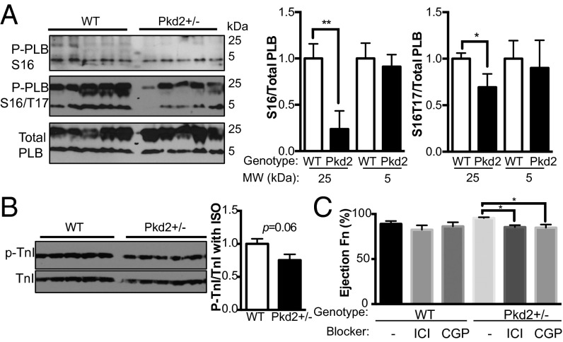
Cardiac disorders are the main cause of mortality in autosomal-dominant polycystic kidney disease (ADPKD). However, how mutated polycystins predispose patients with ADPKD to cardiac pathologies before development of renal dysfunction is unknown. We investigate the effect of decreased levels of polycystin 2 (PC2), a calcium channel that interacts with the ryanodine receptor, on myocardial function. We hypothesize that heterozygous PC2 mice (Pkd2(+/-)) undergo cardiac remodeling as a result of changes in calcium handling, separate from renal complications. We found that Pkd2(+/-) cardiomyocytes have altered calcium handling, independent of desensitized calcium-contraction coupling. Paradoxically, in Pkd2(+/-) mice, protein kinase A (PKA) phosphorylation of phospholamban (PLB) was decreased, whereas PKA phosphorylation of troponin I was increased, explaining the decoupling between calcium signaling and contractility. In silico modeling supported this relationship. Echocardiography measurements showed that Pkd2(+/-) mice have increased left ventricular ejection fraction after stimulation with isoproterenol (ISO), a β-adrenergic receptor (βAR) agonist. Blockers of βAR-1 and βAR-2 inhibited the ISO response in Pkd2(+/-) mice, suggesting that the dephosphorylated state of PLB is primarily by βAR-2 signaling. Importantly, the Pkd2(+/-) mice were normotensive and had no evidence of renal cysts. Our results showed that decreased PC2 levels shifted the βAR pathway balance and changed expression of calcium handling proteins, which resulted in altered cardiac contractility. We propose that PC2 levels in the heart may directly contribute to cardiac remodeling in patients with ADPKD in the absence of renal dysfunction.

Keywords: calcium signaling; excitation contraction coupling; β-adrenergic receptor blocker.


Conflict of interest statement

The authors declare no conflict of interest.

Figures

Fig. 1.
Fig. 1.
Calcium handling in 5-mo-old WT and Pkd2+/− cardiomyocytes. (A) Representative cardiomyocytes loaded with Fura-2AM. (B) Diastolic calcium is unchanged between WT and Pkd2+/− cells. (C) Calcium transients elicited by the addition of 5 mmol/L caffeine to activate release from the RyR-controlled sarcoplasmic reticulum stores from WT (black trace) and Pkd2+/− cardiomyocytes (gray trace). (D) Quantification of area under the curve (AUC) after addition of 5 mmol/L caffeine. Data are the mean ± SEM from n = 9–10 cardiomyocytes, n = 3 animals (*P < 0.05). (E) Magnitude of calcium release (Ca2+ Rmag) is lower in WT (black) than in Pkd2+/− (gray) cardiomyocytes (n = 256 WT, n = 249 Pkd2+/−cells, n = 4 WT, n = 3 Pkd2+/− animals per group; ****P < 0.001). (F) Tau, the rate of calcium decay (TauCa2+), was faster in WT (open bar) than in Pkd2+/− (black bar) cardiomyocytes (n = 256 WT, n = 249 Pkd2+/−cells, n = 4 WT, n = 3 Pkd2+/− animals per group).
Fig. 2.
Fig. 2.
Calcium handling proteins in WT and Pkd2+/− mice. (A) Protein expression of calcium-contractile proteins in WT and Pkd2+/− mice in 5-mo-old mice. Tissue taken from the LV (Left) and right ventricle (Right). Each lane is a separate animal. (B) Quantification of n = 5 WT and n = 4 Pkd2+/− mice normalized to tubulin (LV only; *P < 0.05).
Fig. 3.
Fig. 3.
Pkd2+/− cardiomyocytes have a paradoxical calcium-contractility relationship. (A) Peak sarcomere length shortening (peak SL shortening) in WT (open bar) and Pkd2+/− (black bar) cardiomyocytes is similar. (B) RT50 in WT (open bar) and Pkd2+/− (black bar) cardiomyocytes is similar. (C, Left) Representative traces of experimental cardiomyocyte dynamics for calcium transient traces (Top) from WT (black) and Pkd2+/− (gray) with their corresponding sarcomere length shortening traces (Bottom). (Middle) A computational model whereby the calcium transient for WT cardiomyocytes was used to adjust the shortening traces to match experimental data, then the Pkd2+/− calcium trace was entered and the resultant “expected” sarcomere shortening was recorded. Note that the simulated calcium transient does not match the experimental findings. (Right) A parameter that tunes the rate of calcium dissociation (i.e., TnI phosphorylation) was increased in the Pkd2+/− model, and the output was recorded to match the experimental data. (D) Phosphorylated TnI under baseline conditions in Pkd2+/− mice is higher than in WT mice. For Western blots, each lane represents a different animal (n = 5–6 animals per group) and values are normalized to the total amount of TnI. Tissue is from the LV. WT is represented by open bar, Pkd2+/− by black bar (**P < 0.01).
Fig. 4.
Fig. 4.
PKA phosphorylated proteins localize with the contractile apparatus. (A) TnI (red) and the PKA phosphorylated substrate (green) colocalize. Note that there is a higher degree of colocalization in the Pkd2+/− cardiomyocytes compared with the WT, as quantified to the right. (B) PLB (red) is largely excluded from the contractile apparatus (TnI; green). Images are representative of three mice, and are taken from the LV (*P < 0.05).
Fig. 5.
Fig. 5.
Pkd2+/− mice have enhanced contractility after ISO stimulus. (A) Echocardiograms 3 min after ISO injection (0.1 mg/kg) demonstrate increased LV contractility in 5-mo-old Pkd2+/− mice. (B) No difference in LVEF at baseline between WT and Pkd2+/− mice. (C) LVEF in WT and Pkd2+/− mice after administration of 0.1 mg/kg ISO. Data are presented as mean ± SEM (n = 5–9 animals per group), and values are included in Tables S3 and S4). (D) Blood pressure measurements for 5-mo-old WT and Pkd2+/− mice (n = 5–9 animals per group). WT is represented by open bar, Pkd2+/− by black bar (*P < 0.05).
Fig. 6.
Fig. 6.
Effect of ISO on protein phosphorylation in Pkd2+/− mice and blockade by βAR blockers. (A) Injection of ISO does not induce the same level of phosphorylation of PLB at Ser16 or Ser16/Thr17 in Pkd2+/− mice as in WT mice. Each lane represents a different animal (n = 6 per genotype). (Right) Analysis of data at left. (B) ISO application induces phosphorylation of TnI in tissue from ISO-treated animals. (Right) Analysis of data at left. WT is represented by open bar, Pkd2+/− by black bar. (C) Effect of the β-blockers ICI 118,551 and CGP 20712 on WT and Pkd2+/− 5-mo-old mice 3 min after ISO application (n = 4–6 animals per group; *P < 0.05 and **P < 0.01).

Similar articles

Cited by

References

    1. Chapman AB, Stepniakowski K, Rahbari-Oskoui F. Hypertension in autosomal dominant polycystic kidney disease. Adv Chronic Kidney Dis. 2010;17(2):153–163. - PMC - PubMed
    1. Ecder T, Schrier RW. Cardiovascular abnormalities in autosomal-dominant polycystic kidney disease. Nat Rev Nephrol. 2009;5(4):221–228. - PMC - PubMed
    1. Schrier RW. Renal volume, renin-angiotensin-aldosterone system, hypertension, and left ventricular hypertrophy in patients with autosomal dominant polycystic kidney disease. J Am Soc Nephrol. 2009;20(9):1888–1893. - PubMed
    1. Bardají A, et al. Cardiac involvement in autosomal-dominant polycystic kidney disease: a hypertensive heart disease. Clin Nephrol. 2001;56(3):211–220. - PubMed
    1. Cadnapaphornchai MA, McFann K, Strain JD, Masoumi A, Schrier RW. Increased left ventricular mass in children with autosomal dominant polycystic kidney disease and borderline hypertension. Kidney Int. 2008;74(9):1192–1196. - PMC - PubMed

Publication types

MeSH terms

LinkOut - more resources