. 2014 Dec 26;289(52):35695-710.
doi: 10.1074/jbc.M114.584284. Epub 2014 Nov 3.

Syk and Src family kinases regulate C-type lectin receptor 2 (CLEC-2)-mediated clustering of podoplanin and platelet adhesion to lymphatic endothelial cells

Affiliations

Syk and Src family kinases regulate C-type lectin receptor 2 (CLEC-2)-mediated clustering of podoplanin and platelet adhesion to lymphatic endothelial cells

Alice Y Pollitt et al. J Biol Chem. .

Abstract

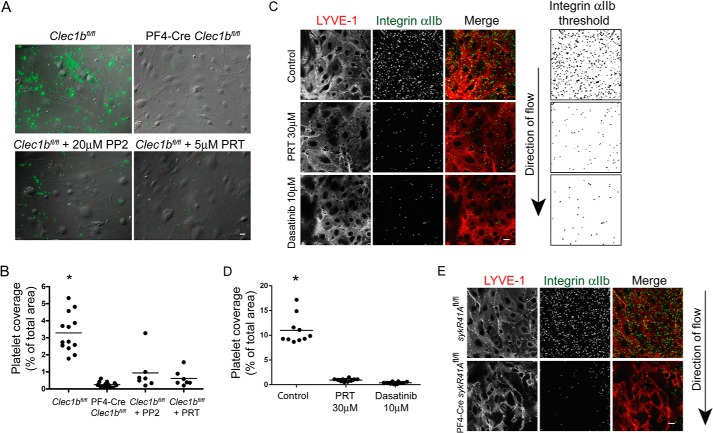
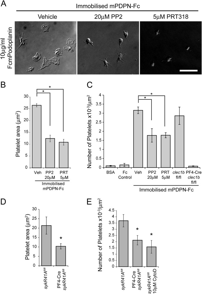
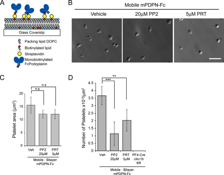
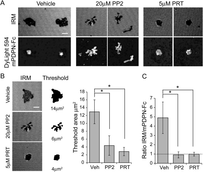
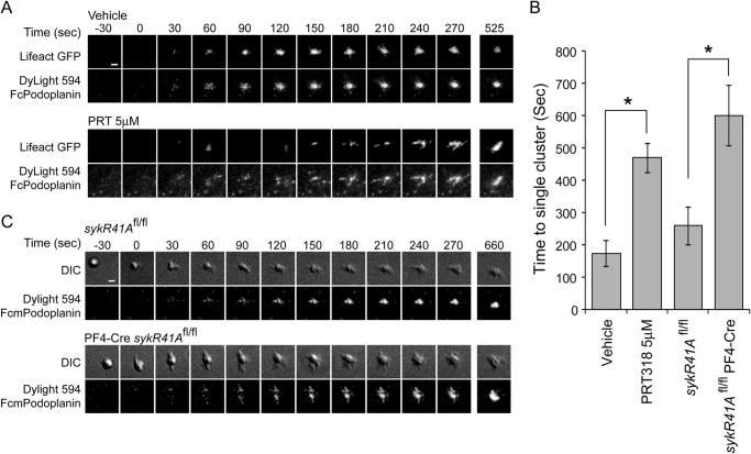
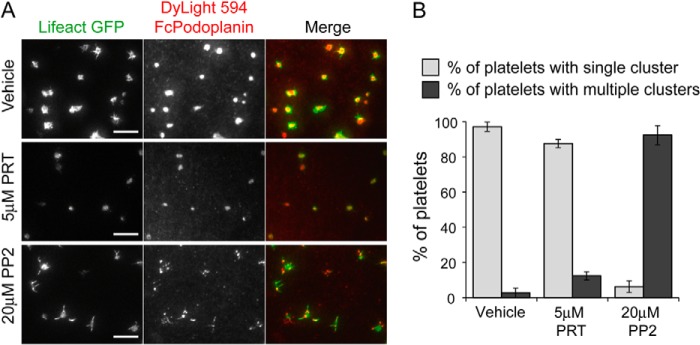
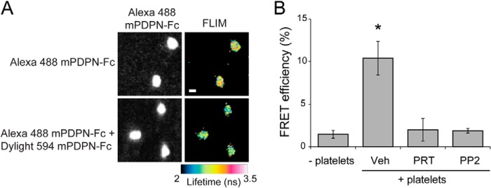
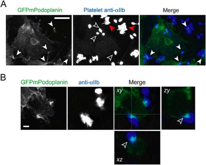
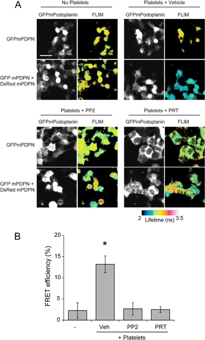
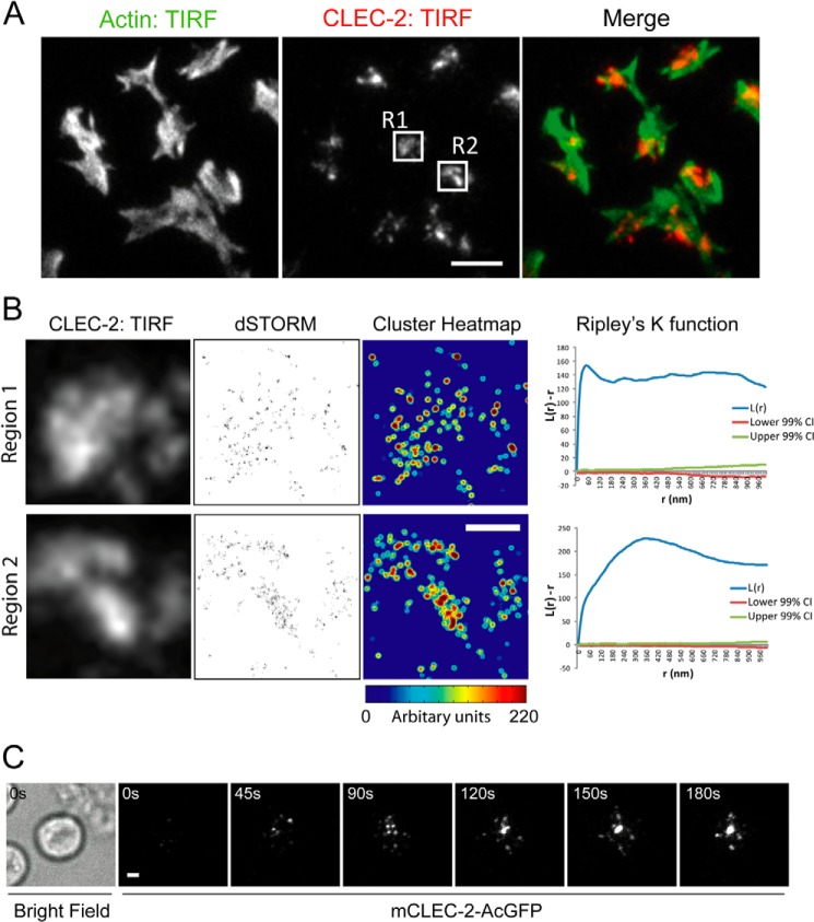
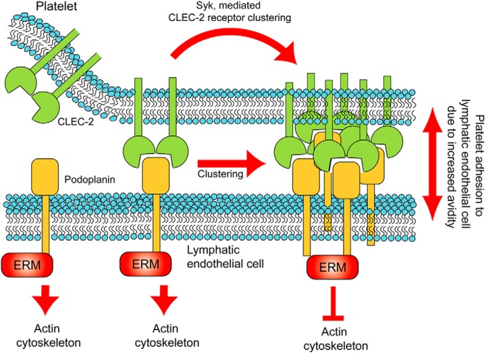
The interaction of C-type lectin receptor 2 (CLEC-2) on platelets with Podoplanin on lymphatic endothelial cells initiates platelet signaling events that are necessary for prevention of blood-lymph mixing during development. In the present study, we show that CLEC-2 signaling via Src family and Syk tyrosine kinases promotes platelet adhesion to primary mouse lymphatic endothelial cells at low shear. Using supported lipid bilayers containing mobile Podoplanin, we further show that activation of Src and Syk in platelets promotes clustering of CLEC-2 and Podoplanin. Clusters of CLEC-2-bound Podoplanin migrate rapidly to the center of the platelet to form a single structure. Fluorescence lifetime imaging demonstrates that molecules within these clusters are within 10 nm of one another and that the clusters are disrupted by inhibition of Src and Syk family kinases. CLEC-2 clusters are also seen in platelets adhered to immobilized Podoplanin using direct stochastic optical reconstruction microscopy. These findings provide mechanistic insight by which CLEC-2 signaling promotes adhesion to Podoplanin and regulation of Podoplanin signaling, thereby contributing to lymphatic vasculature development.

Keywords: CLEC-2; Endothelial Cell; ITAM; Lipid Bilayer; Platelet; Podoplanin; Receptor; Src Family Kinase; Syk; Tyrosine-Protein Kinase (Tyrosine Kinase).


Figures

FIGURE 1.
FIGURE 1.
CLEC-2 signaling supports platelet adhesion to primary mouse lymphatic endothelial cells under static and flow conditions. A, CLEC-2-deficient (PF4-Cre Clec1bfl/fl) or control (Clec1bfl/fl) platelets in the presence or absence of inhibitors to Src family (PP2, 20 μm) or Syk (PRT-060318, 5 μm) kinases were incubated with a confluent monolayer of primary mouse LECs (DIC image) for 1 h at 37 °C. Platelets were identified using and anti-αIIb antibody (green). Scale bar, 20 μm. B, quantification of CLEC-2-deficient (PF4-CreClec1bfl/fl) or control (Clec1bfl/fl) platelet coverage in the presence or absence of inhibitors to Src family (PP2, 20 μm) or Syk (PRT-060318, 5 μm) kinases. *, p < 0.01 in analysis of variance. C, control blood in the presence or absence of inhibitors to Src family (10 μm dasatinib) or Syk (PRT-060318, 30 μm) kinases was perfused over primary mouse LECs at a wall shear rate of 50 s−1 at 37 °C. An arrow indicates the direction of flow. LECs and platelets were identified using anti-LYVE-1 and anti-αIIb antibodies, respectively. Integrin αIIb antibody threshold (right panels). Scale bar, 20 μm. D, quantification of platelet adhesion to primary mouse LECs under flow conditions in the presence or absence of inhibitors to Src family kinases (10 μm dasatinib) or Syk kinase (PRT-060318, 30 μm). E, control (SykR41A fl/fl) or SykR41A expressing (PF4-CreSykR41Afl/fl) mouse blood was perfused over primary mouse LECs at a wall shear rate of 50 s−1 at 37 °C. An arrow indicates the direction of flow. LECs and platelets were identified using anti-LYVE-1 and anti-αIIb antibodies, respectively. Scale bar, 20 μm. *, p < 0.01 in analysis of variance.
FIGURE 2.
FIGURE 2.
Syk and Src family kinases support platelet adhesion to immobilized Podoplanin. A, washed platelets in the presence or absence of Src family (PP2, 20 μm) or Syk (PRT-060318, 5 μm) kinase inhibitors were allowed to interact with mPDPN-Fc (10 μg/ml)-coated glass coverslips for 45 min at 37 °C and imaged using DIC microscopy. Scale bar, 10 μm. B, quantification of platelet area when interacting with immobilized mPDPN-Fc in the presence or absence of inhibitors to Src family (PP2, 20 μm) or Syk (PRT-060318, 5 μm) kinases. C, quantification of the number of adherent platelets interacting with BSA, a control Fc protein, or immobilized mPDPN-Fc with or without Src family (PP2, 20 μm) or Syk (PRT-060318, 5 μm) kinase inhibitors. Quantification of the number of CLEC-2-deficient (PF4-Cre Clec1bfl/fl) or control (Clec1bfl/fl) platelets interacting with mPDPN-Fc. D, quantification of control (SykR41A fl/fl) or SykR41A expressing (PF4-CreSykR41Afl/fl) platelet area when interacting with immobilized mPDPN-Fc. E, quantification of the number of adhered SykR41A expressing (PF4-CreSykR41Afl/fl) or control (SykR4A fl/fl) platelets with or without 10 μm cytochalasin D interacting with immobilized mPDPN-Fc. Images and results are the averages of three independent experiments ± S.D. *, p < 0.05. Veh, vehicle.
FIGURE 3.
FIGURE 3.
Syk and Src family kinases support platelet adhesion to mobile Podoplanin. A, schematic of the experimental system for mobile Podoplanin. Platelets were allowed to interact with glass-supported planar lipid bilayers containing biotinylated lipids, to which monobiotinylated recombinant Podoplanin (mPDPN-Fc) was tethered via streptavidin. DOPC, dioleoylphosphocholine. B, washed platelets pretreated with or without Src family (PP2, 20 μm) or Syk (PRT-060318, 5 μm) kinase inhibitors were allowed to interact with glass-supported planar lipid bilayers containing mobile mPDPN-Fc for 45 min at 37 °C and imaged using DIC microscopy. Scale bar, 10 μm. C, quantification of platelet area when interacting with mobile mPDPN-Fc with or without inhibitors to Src family (PP2, 20 μm) or Syk (PRT-060318, 5 μm) kinases. The results are the averages of four independent experiments ± S.D. D, quantification of the number of adherent platelets interacting with mobile mPDPN-Fc with or without inhibitors to Src family (PP2, 20 μm) or Syk (PRT-060318, 5 μm) kinases. Images and results are the averages of three independent experiments ± S.D. **, p < 0.01; ***, p < 0.001. n.s., not significant; Veh, vehicle.
FIGURE 4.
FIGURE 4.
Syk and Src family kinase inhibition decreases platelet-bilayer interactions. A, platelets treated with vehicle, Syk (PRT-060318, 5 μm) or Src family (PP2 20 μm) kinase inhibitors were allowed to interact with glass-supported lipid bilayers containing Dylight 594-labeled mPDPN-Fc for 45 min at 37 °C. IRM and fluorescent images of the platelet/bilayer interface. Top panels, IRM; bottom panels, confocal images of Dylight 594-labeled mPDPN-Fc. B, example platelet contact area shown as the original unthresholded image (left panels) or after thresholding and binarization (right panels). Quantification of close surface contact areas of platelets interacting with mobile Podoplanin is shown. C, the ratio of the IRM threshold area to mPDPN-Fc area. The results are the averages of three separate experiments ± S.D. *, p < 0.05. Veh, vehicle.
FIGURE 5.
FIGURE 5.
Role of Syk kinase in platelet-mediated Podoplanin cluster dynamics. A, TIRFM time course of Lifeact-GFP-expressing platelets interacting with planar lipid bilayer containing Dylight 594-labeled mPDPN-Fc. The top panels show control Lifeact-GFP-expressing platelets. The bottom panels show Lifeact-GFP platelets in the presence of a Syk kinase inhibitor (PRT-060318, 5 μm; see supplemental Movie S1). B, quantification of the time taken to form a single stable central structure from the first point of contact. The results are the averages of three separate experiments ± S.D. *, p < 0.01. C, DIC time course of control (sykR41Afl/fl; upper panels) or SykR41A expressing (PR4-CresykR41Afl/fl; lower panels) platelets interacting with planar lipid bilayer containing Dylight 594-labeled mPDPN-Fc (see supplemental Movie S2). Time is relative to the first detected contact point. Scale bars, 2 μm.
FIGURE 6.
FIGURE 6.
Src family kinase inhibition blocks Podoplanin cluster dynamics. A, TIRFM images of Lifeact-GFP-expressing platelets interacting with planar lipid bilayer containing Dylight 594-labeled mPDPN-Fc for 45 min at 37 °C. Lifeact-GFP-expressing platelets were pretreated with Src family kinase inhibitor (PP2, 20 μm) or a Syk inhibitor (PRT-060318, 5 μm). Scale bar, 10 μm. B, quantification of the cluster morphology in the presence and absence of Src family (PP2, 20 μm) or a Syk (PRT-060318, 5 μm) kinase inhibitors at 45 min. Platelets were identified as having a single or multiple Podoplanin clusters. Images and results are the averages of three independent experiments ± S.D.
FIGURE 7.
FIGURE 7.
Syk and Src family kinase inhibition decreases Podoplanin clustering. A, platelets interacting with planar lipid bilayer containing Alexa 488-labeled mPDPN-Fc or equivalent amounts of Alexa 488-labeled mPDPN-Fc and Dylight 594-labeled mPDPN-Fc for 45 min at 37 °C. Fluorescence lifetimes of the donor fluorophore (mPDPN-Fc-488) were determined in the absence and presence of acceptor fluorophore (mPDPN-Fc-594). The fluorescence lifetimes in nanoseconds (ns) are shown as false color images. Scale bar, 2 μm. B, FRET-FLIM analysis of mPDPN-Fc containing bilayers incubated with platelets treated with or without inhibitors to Src family (PP2, 20 μm) or Syk (PRT-060318, 5 μm) kinases. Images and results are the averages of three independent experiments ± S.D. Veh, vehicle.
FIGURE 8.
FIGURE 8.
Podoplanin is recruited at the platelet-cell interface. A, HEK293T cells expressing GFPmPodoplanin were analyzed in the presence of platelets by confocal microscopy. Maximum intensity projection of GFPmPodoplanin HEK293T cells (left panel) and platelets stained with an anti-αIIb antibody (middle panel). The merged images are shown in the right panel (GFPmPodoplanin, green; anti-αIIb, blue). Clusters of GFPmPodoplanin associated with a platelet are indicated by closed white arrowheads in the GFPmPodoplanin image and superimposed on the merge image. Platelets only interacting with the GFPmPodoplanin HEK293T cell are indicated by open arrowheads. Platelets interacting with both the GFPmPodoplanin HEK293T cell and the coverslip are indicated by red arrowheads. Scale bar, 10 μm. B, confocal image of GFPmPodoplanin (left panel) and anti-αIIb antibody (middle panel). The merged images are shown in the right panel (GFPmPodoplanin, green; anti-αIIb, Blue). xz and yz projections display a cross-section of a cell-platelet interaction. The accumulation of GFPmPodoplanin between the cell and the platelet is indicated by an open arrowhead in the xz and yz views. Scale bar, 2 μm. Images are representative of three independent experiments.
FIGURE 9.
FIGURE 9.
CLEC-2 signaling leads to Podoplanin clustering in a cell membrane. A, platelets were allowed to interact with cells expressing either AcGFPmPodoplanin or cells expressing equivalent levels of AcGFPmPodoplanin and DsRedmonomer-mPodoplanin for 1 h. Samples were fixed, and the average lifetime of AcGFPmPodoplanin was determined in the absence and presence of the acceptor DsRedmPodoplanin for cells incubated with platelets treated with vehicle, Syk (PRT-060318, 5 μm), or Src family (PP2 20 μm) kinase inhibitors. The fluorescence lifetimes in nanoseconds (ns) are shown as false color images. Scale bar, 10 μm. B, FRET-FLIM analysis of cells expressing AcGFPmPodoplanin incubated with platelets incubated with or without Syk (PRT-060318, 5 μm) or Src family (PP2 20 μm) kinase inhibitors. Images and results are the averages of three separate experiments ± S.D. *, p < 0.01. Veh, vehicle.
FIGURE 10.
FIGURE 10.
CLEC-2 can form clusters that, following ligand engagement, migrate in a directed manner toward the center of the cell. A, washed mouse platelets were allowed to interact with mPDPN-Fc (10 μg/ml)-coated glass dishes for 45 min at 37 °C. TIRFM image of Alexa 488-phalloidin (left panel) and Alexa 647-anti-CLEC-2 (INU1) (middle panel). The merged images are shown in the right panel (actin, green; mCLEC-2, red). Two regions of interest are highlighted, R1 and R2. Scale bar, 5 μm. B, TIRFM images of Alexa 647-anti-mCLEC-2 in the boxed regions from Fig. 10A (R1 and R2; left panels). dSTORM images of regions 1 and 2 (middle panels). Quantitative cluster mapping of the dSTORM images of regions 1 and 2 (right panels). Scale bar, 1 μm. Ripley's K-function analysis of the molecules in regions 1 and 2; L(r) − r reports the degree of clustering relative to a random distribution (indicated by the lower and upper 99% confidence intervals (CI) (red and green lines)), and r is the radial scale. C, TIRFM time course of a mCLEC-2-AcGFP-expressing DT40 chicken B cell interacting with a planar lipid bilayer containing an CLEC-2-activating antibody (17D9) (see supplemental Movie S3). The bright field image shows the location of the cell. Scale bar, 2 μm. Images are representative of three independent experiments.
FIGURE 11.
FIGURE 11.
Model summary of CLEC-2-mediated clustering of Podoplanin. We propose that the binding of CLEC-2 to Podoplanin enhances the adhesion of platelets to lymphatic endothelial cells through CLEC-2-mediated receptor clustering involving the kinase Syk. We hypothesize that this would in turn lead to clustering of Podoplanin and functional effects in the LECs by modulation of Podoplanin signaling though ERM proteins and regulation of the actin cytoskeleton.

References

    1. Suzuki-Inoue K., Fuller G. L., García A., Eble J. A., Pöhlmann S., Inoue O., Gartner T. K., Hughan S. C., Pearce A. C., Laing G. D., Theakston R. D., Schweighoffer E., Zitzmann N., Morita T., Tybulewicz V. L., Ozaki Y., Watson S. P. (2006) A novel Syk-dependent mechanism of platelet activation by the C-type lectin receptor CLEC-2. Blood 107, 542–549 - PubMed
    1. Colonna M., Samaridis J., Angman L. (2000) Molecular characterization of two novel C-type lectin-like receptors, one of which is selectively expressed in human dendritic cells. Eur. J. Immunol. 30, 697–704 - PubMed
    1. Séverin S., Pollitt A. Y., Navarro-Nuñez L., Nash C. A., Mourão-Sá D., Eble J. A., Senis Y. A., Watson S. P. (2011) Syk-dependent phosphorylation of CLEC-2: a novel mechanism of hem-immunoreceptor tyrosine-based activation motif signaling. J. Biol. Chem. 286, 4107–4116 - PMC - PubMed
    1. Spalton J. C., Mori J., Pollitt A. Y., Hughes C. E., Eble J. A., Watson S. P. (2009) The novel Syk inhibitor R406 reveals mechanistic differences in the initiation of GPVI and CLEC-2 signaling in platelets. J. Thromb. Haemost. 7, 1192–1199 - PubMed
    1. Hughes C. E., Sinha U., Pandey A., Eble J. A., O'Callaghan C. A., Watson S. P. (2013) Critical Role for an acidic amino acid region in platelet signaling by the HemITAM (hemi-immunoreceptor tyrosine-based activation motif) containing receptor CLEC-2 (C-type lectin receptor-2). J. Biol. Chem. 288, 5127–5135 - PMC - PubMed

Publication types