. 2015 Mar;13(3):502-9.
doi: 10.1158/1541-7786.MCR-14-0407. Epub 2014 Nov 3.

The Tyrosine Kinase Adaptor Protein FRS2 Is Oncogenic and Amplified in High-Grade Serous Ovarian Cancer

Affiliations

The Tyrosine Kinase Adaptor Protein FRS2 Is Oncogenic and Amplified in High-Grade Serous Ovarian Cancer

Leo Y Luo et al. Mol Cancer Res. 2015 Mar.

Abstract

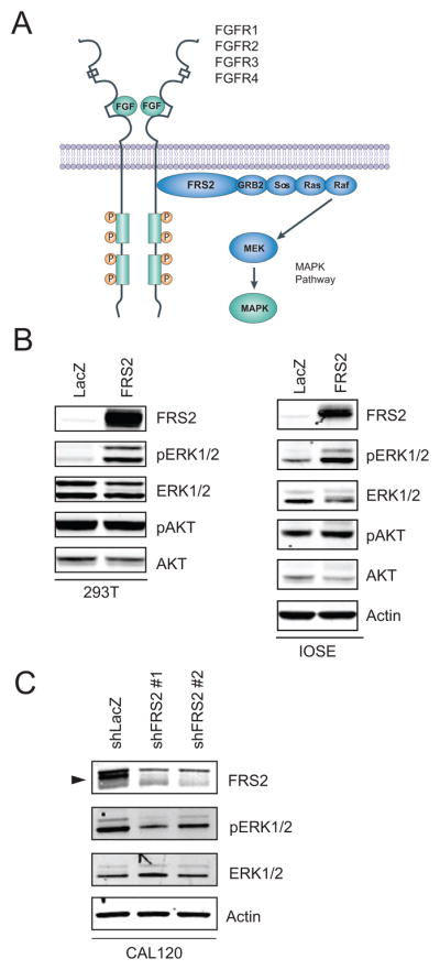
High-grade serous ovarian cancers (HGSOC) are characterized by widespread recurrent regions of copy-number gain and loss. Here, we interrogated 50 genes that are recurrently amplified in HGSOC and essential for cancer proliferation and survival in ovarian cancer cell lines. FRS2 is one of the 50 genes located on chromosomal region 12q15 that is focally amplified in 12.5% of HGSOC. We found that FRS2-amplified cancer cell lines are dependent on FRS2 expression, and that FRS2 overexpression in immortalized human cell lines conferred the ability to grow in an anchorage-independent manner and as tumors in immunodeficient mice. FRS2, an adaptor protein in the FGFR pathway, induces downstream activation of the Ras-MAPK pathway. These observations identify FRS2 as an oncogene in a subset of HGSOC that harbor FRS2 amplifications.

Implications: These studies identify FRS2 as an amplified oncogene in a subset of HGSOC. FRS2 expression is essential to ovarian cancer cells that harbor 12q15 amplification.


Conflict of interest statement

Disclosure of Potential Conflicts of Interest: WCH is a consultant to Novartis and RRS is an employee of Astellas Pharma US.

Figures

Figure 1
Figure 1. Amplification and Overexpression of FRS2 in primary high-grade serous ovarian cancers and ovarian cancer cell lines
A. FRS2 is one of the 50 genes that are recurrently amplified in primary ovarian tumors and essential for ovarian cancer cell proliferation and survival. B. Copy number profile along chromosome 12q of human tumor samples. FRS2 was amplified in multiple cancer types including ovarian, breast, lung squamous, lung adenocarcinoma, stomach, head and neck (H&N), and bladder. Each vertical line represents one tumor sample. Red represents copy number gain, Blue represents copy number loss. C. Level of FRS2 mRNA expression in primary tumors correlates with the copy number. Copy number is divided into 4 categories based on log2 of copy numbers. “Amplification” is defined as Log2(Copy number) more than 1; “Gain” is between 0.2 and 1; “Normal” is between −0.2 and 0.2; “Loss” is less than −0.2. D. FRS2 amplification and FGFR1, FGFR2, FGFR3, and FGFR4 amplifications are mutually exclusive in high-grade serous ovarian cancers. Data was analyzed using the cBio portal rather than GISTIC.
Figure 2
Figure 2. Suppression of FRS2 decreases the proliferation of ovarian and breast cancer cells harboring 12q15 amplification
A. SNP array colorgram showing genomic amplification of chromosome 12q15 in ovarian and breast cancer cell lines. Red represents copy number amplification, blue represents copy number deletion. B. Consequences of FRS2 suppression on the proliferation of cancer cell line that either harbor 12q15 amplification (CAL120, HCC1143, COV644) or normal copy number of 12q15 (CAOV3, COV362, EFO21) normalized to cells treated with shLacZ. Red: cell lines treated shFRS2 #1. Black: cell lines treated with shFRS2#2. **P < 0.01 compared to control shLacZ, Student’s t test was used. C. Quantitative RT-PCR of FRS2 expression in FRS2 amplified (red) and non-amplified (black) cell lines. D. Increased apoptosis in FRS2 amplified cell lines (red) upon FRS2 suppression, shown by increased PARP cleavage.
Figure 3
Figure 3. FRS2 overexpression potentiates tumorigenicity
A. FRS2 promotes anchorage-independent growth in HA1E-A cells compared to LacZ control. MEK-DD, a constitutively active MEK, is positive control. Right, images of soft agar colonies formed by HA1E-A with either FRS2 or control vector overexpression. B. FRS2 promotes anchorage independent growth of IOSE (immortalized human ovarian epithelial) cells. GAB2 is a similar adaptor protein known to transform ovarian epithelial cells. **P < 0.01, ***P < 0.001 compared to respective control vectors, Student’s t test. C. FRS2 overexpression promotes tumorigenicity in vivo. 3T3 cells with FRS2 overexpression were able to form tumor in mouse xenograft models compared to LacZ control. Constitutively active KRAS G13D was used as positive control. Within each condition, tumors from the same mouse were annotated with same color.
Figure 4
Figure 4. FRS2 promotes tumorigenesis via activation of MAPK pathway
A. FRS2 functions as an adaptor protein in the fibroblast growth factor receptor signaling pathway, adapted from Turner and Grose (40). B. Effect of FRS2 overexpression on phosphorylation of ERK in 293T cells and ovarian epithelial cells. C. Effect of FRS2 suppression on phosphorylation of ERK in cancer cell line with 12q15 amplification.

Similar articles

Cited by

References

    1. Bowtell DD. The genesis and evolution of high-grade serous ovarian cancer. Nat Rev Cancer. 2010;10:803–8. - PubMed
    1. Bell D, Berchuck A, Birrer M, Chien J, Cramer DW, Dao F, et al. Integrated genomic analyses of ovarian carcinoma. Nature. 2011;474:609–15. - PMC - PubMed
    1. Cheung HW, Cowley GS, Weir BA, Boehm JS, Rusin S, Scott JA, et al. Systematic investigation of genetic vulnerabilities across cancer cell lines reveals lineage-specific dependencies in ovarian cancer. P Natl Acad Sci USA. 2011;108:12372–7. - PMC - PubMed
    1. Luo B, Cheung HW, Subramanian A, Sharifnia T, Okamoto M, Yang XP, et al. Highly parallel identification of essential genes in cancer cells. P Natl Acad Sci USA. 2008;105:20380–5. - PMC - PubMed
    1. Gao J, Aksoy BA, Dogrusoz U, Dresdner G, Gross B, Sumer SO, et al. Integrative analysis of complex cancer genomics and clinical profiles using the cBioPortal. Sci Signal. 2013;6:pl1. - PMC - PubMed

Publication types

MeSH terms

Substances