. 2014 Oct 16;442(17):132-136.
doi: 10.1016/j.chemphys.2014.02.018.

The enzymatic reaction catalyzed by lactate dehydrogenase exhibits one dominant reaction path

Affiliations

The enzymatic reaction catalyzed by lactate dehydrogenase exhibits one dominant reaction path

Jean E Masterson et al. Chem Phys. .

Abstract

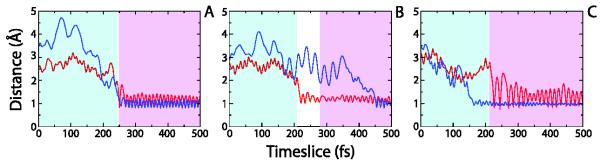
Enzymes are the most efficient chemical catalysts known, but the exact nature of chemical barrier crossing in enzymes is not fully understood. Application of transition state theory to enzymatic reactions indicates that the rates of all possible reaction paths, weighted by their relative probabilities, must be considered in order to achieve an accurate calculation of the overall rate. Previous studies in our group have shown a single mechanism for enzymatic barrier passage in human heart lactate dehydrogenase (LDH). To ensure that this result was not due to our methodology insufficiently sampling reactive phase space, we implement high-perturbation transition path sampling in both microcanonical and canonical regimes for the reaction catalyzed by human heart LDH. We find that, although multiple, distinct paths through reactive phase space are possible for this enzymatic reaction, one specific reaction path is dominant. Since the frequency of these paths in a canonical ensemble is inversely proportional to the free energy barriers separating them from other regions of phase space, we conclude that the rarer reaction paths are likely to have a negligible contribution. Furthermore, the non-dominate reaction paths correspond to altered reactive conformations and only occur after multiple steps of high perturbation, suggesting that these paths may be the result of non-biologically significant changes to the structure of the enzymatic active site.

Keywords: enzymatic catalysis; lactate dehydrogenase; reaction coordinate; transition path sampling.


Figures

Figure 1
Figure 1
Schematic of the reaction catalyzed by human heart lactate dehydrogenase shown from the side of pyruvate. The hydride and proton transfers are depicted in red and blue, respectively. Arginine 169 coordinates the substrate in the active site, and arginine 106 acts to polarize the bond of the substrate keto group.
Figure 2
Figure 2
Progression of a TPE through reactive phase space. This graph shows all 50 reactive trajectories of an example TPE (cTPE1). The y-dimension is the time length of each trajectory (500 timeslices of 1 fs each), and the x-dimension is the trajectory number within the ensemble. The solid and dashed black lines follow along the points of hydride and proton transfer (respectively) for each trajectory. The blue region represents Type 1 trajectories, the orange region contains Type 2 trajectories, and the red region corresponds to Type 3 trajectories.
Figure 3
Figure 3
Example trajectories for each LDH mechanism. Dynamic distances between the particles and the particle acceptors are red lines for the hydride and blue lines for the proton. Cyan regions correspond to the pyruvate basins, and the lavender regions represent the lactate basins. (A) Type 1 mechanism with a barrier crossing of 3 fs, (B) Type 2 mechanism with a barrier crossing of 78 fs, and (C) Type 3 mechanism with a barrier crossing of 3 fs.
Figure 4
Figure 4
Transition state formation for each LDH mechanism. Example trajectories of (A) a Type 1 mechanism, (B) a Type 2 mechanism, and (C) a Type 3 mechanism are shown as graphs. The gray lines represent the transition state as determined by committor analysis, red dashed lines are the hydride donor—acceptor distances, and blue dashed lines are the proton donor— acceptor distances. Schematics of the transition states for each mechanism are explicitly shown with the hydride—acceptor bond in red and the proton—acceptor bond in blue.
Figure 5
Figure 5
Reactive conformations of LDH. The black squares represent the first 50 reactive trajectories of a TPE previously generated using standard sampling methods, all of which display a Type 1 mechanism. The circles correspond to 50 reactive trajectories comprising a TPE generated via high-perturbation TPS (cTPE1). Blue circles represent trajectories which exhibited a Type 1 mechanism, whereas orange circles are trajectories that display a Type 2 mechanism and red circles are trajectories that react via a Type 3 mechanism.

References

    1. Antoniou D, Schwartz SD. Protein dynamics and enzymatic chemical barrier passage. J. Phys. Chem. B. 115(2011):15147–15158. - PMC - PubMed
    1. Schwartz SD, Schramm VL. Enzymatic transition states and dynamic motion in barrier crossing. Nature Chem. Biol. 2009;5:551–558. - PMC - PubMed
    1. Herschlag D, Natarajan A. Fundamental challenges in mechanistic enzymology: progress toward understanding the rate enhancements of enzymes. Biochemistry. 2013;52:2050–2067. - PMC - PubMed
    1. Pauling L. Chemical achievement and hope for the future. Am. Sci. 1948;36:51–58. - PubMed
    1. Schultz PG. Catalytic Antibodies. Angew. Chem. Int. Ed. 1989;28:1283–1295.

LinkOut - more resources