. 2014 Oct 17:8:845.
doi: 10.3389/fnhum.2014.00845. eCollection 2014.

Regional white matter damage predicts speech fluency in chronic post-stroke aphasia

Affiliations

Regional white matter damage predicts speech fluency in chronic post-stroke aphasia

Alexandra Basilakos et al. Front Hum Neurosci. .

Abstract

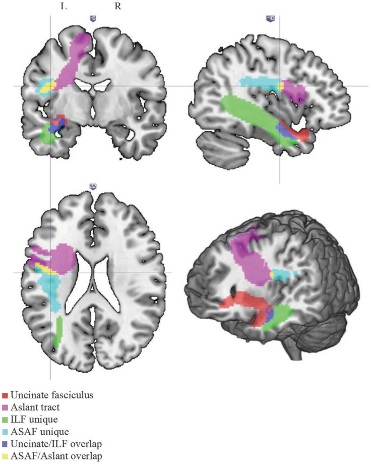
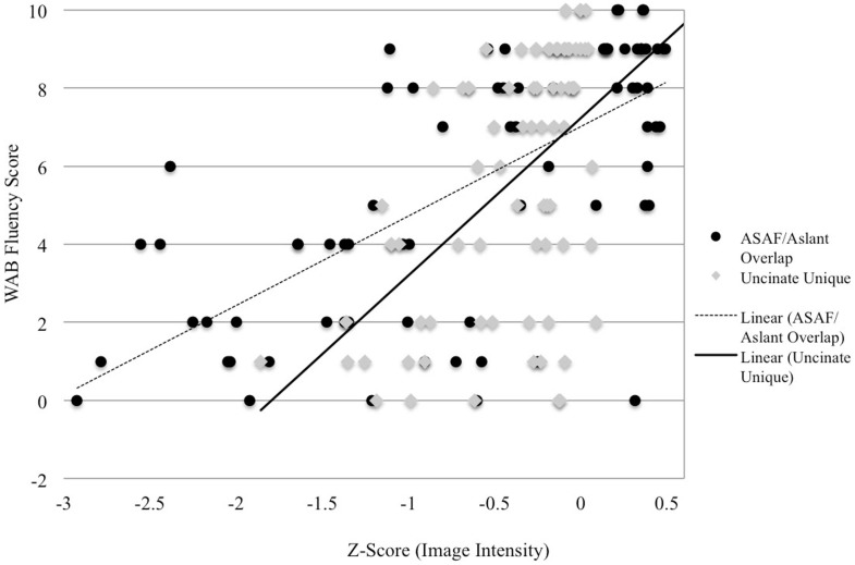
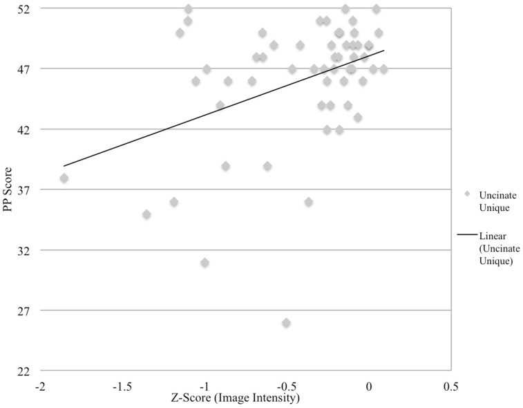
RECENTLY, TWO DIFFERENT WHITE MATTER REGIONS THAT SUPPORT SPEECH FLUENCY HAVE BEEN IDENTIFIED: the aslant tract and the anterior segment of the arcuate fasciculus (ASAF). The role of the ASAF was demonstrated in patients with post-stroke aphasia, while the role of the aslant tract shown in primary progressive aphasia. Regional white matter integrity appears to be crucial for speech production; however, the degree that each region exerts an independent influence on speech fluency is unclear. Furthermore, it is not yet defined if damage to both white matter regions influences speech in the context of the same neural mechanism (stroke-induced aphasia). This study assessed the relationship between speech fluency and quantitative integrity of the aslant region and the ASAF. It also explored the relationship between speech fluency and other white matter regions underlying classic cortical language areas such as the uncinate fasciculus and the inferior longitudinal fasciculus (ILF). Damage to these regions, except the ILF, was associated with speech fluency, suggesting synergistic association of these regions with speech fluency in post-stroke aphasia. These observations support the theory that speech fluency requires the complex, orchestrated activity between a network of pre-motor, secondary, and tertiary associative cortices, supported in turn by regional white matter integrity.

Keywords: aphasia; arcuate fasciculus; frontal aslant tract; inferior longitudinal fasciculus; non-fluent speech; speech production; uncinate fasciculus.


Figures

Figure 1
Figure 1
White matter tracts. Locations of each of the white matter tracts included in our analyses. Note the following color scheme: magenta – aslant tract; cyan – ASAF; red – uncinate fasciculus; yellow – ASAF/aslant overlap; green – ILF; blue – uncinate/ILF overlap. The ASAF forms a connection between the pars opercularis and lateral middle and inferior precentral gyri to the posterior, inferior parietal lobe. The aslant tract connects Broca’s area to pre-supplementary motor areas. The ASAF/aslant overlap underlies BA 6. The uncinate fasciculus connects the anterior and superior temporal lobe to the medial and lateral portions of the inferior frontal cortex. The ILF connects occipitotemporal regions to the temporal pole. The uncinate and ILF overlap in white matter of the inferior temporal lobe (Brodmann area 20) and parahippocampal cortex (Brodmann area 36).
Figure 2
Figure 2
Image intensity and WAB-R fluency scores. Z-scores for T1-MRI image intensity values for the aslant tract and ASAF (x-axis) and WAB-R fluency scores (y-axis). Solid and dotted lines depict regression lines of best fit for the uncinate fasciculus and ASAF/aslant overlap, respectively.
Figure 3
Figure 3
Image intensity and semantic processing. Z-scores for T1-MRI image intensity values for the uncinate fasciculus (x-axis) and pyramids and palms scores (y-axis). Solid line depicts regression line of best.

References

    1. Albert M., Goodglass H., Helm N. A., Rubens A., Alexander M. (1981). Clinical Aspects of Dysphasia. New York, NY: Springer-Verlag
    1. Alexander M. P., Naeser M. A., Palumbo C. (1990). Broca’s area aphasias: aphasia after lesions including the frontal operculum. Neurology 40, 353–362.10.1212/WNL.40.2.353 - DOI - PubMed
    1. Andersen S. M., Rapcsak S. Z., Beeson P. M. (2010). Cost function masking during normalization of brains with focal lesions: still a necessity? Neuroimage 53, 78–84.10.1016/j.neuroimage.2010.06.003 - DOI - PMC - PubMed
    1. Bates E., Wilson S. M., Saygin A. P., Dick F., Sereno M. I., Knight R. T., et al. (2003). Assessing brain-behavior relationships using voxel-based lesion-symptom mapping. Nat. Neurosci. 6, 448–450.10.1038/nn1050 - DOI - PubMed
    1. Boddaert N., Brunelle F., Desquerre I. (2013). Clinical and imaging diagnosis for heredodegenerative diseases. Handb. Clin. Neurol. 111, 63–78.10.1016/B978-0-444-52891-9.00006-3 - DOI - PubMed

LinkOut - more resources