Idiopathic pleuroparenchymal fibroelastosis presenting in recurrent pneumothorax: a case report
- PMID: 25368665
- PMCID: PMC4217035
- DOI: 10.4046/trd.2014.77.4.184
Idiopathic pleuroparenchymal fibroelastosis presenting in recurrent pneumothorax: a case report
Abstract
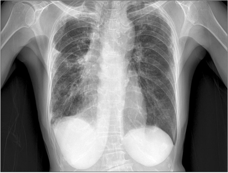
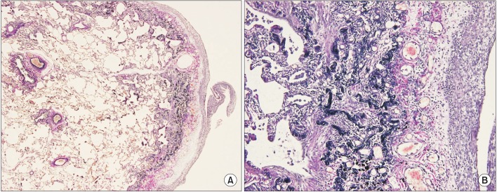
Idiopathic pleuroparenchymal fibroelastosis (PPFE) is a rare, recently classified entity that consists of pleural and subjacent parenchymal fibrosis predominantly in the upper lungs. In an official American Thoracic Society/European Respiratory Society statement in 2013, this disease is introduced as a group of rare idiopathic interstitial pneumonias. We describe a case of a 76-year-old woman with cough and recurrent pneumothorax. She was admitted to our hospital with severe cough at first. High resolution computed tomography (HRCT) disclosed multifocal subpleural consolidations with reticular opacities in both lungs, primarily in the upper lobes, suggesting interstitial pneumonia. Rheumatoid lung was diagnosed initially through an elevated rheumatoid factor, HRCT and surgical biopsy at the right lower lobe. However, one month later, pneumothorax recurred. Surgical biopsy was performed at the right upper lobe at this time. The specimens revealed typical subpleural fibroelastosis. We report this as a first case of idiopathic PPFE in Korea after reviewing the symptoms, imaging and pathologic findings.
Keywords: Fibrosis; Idiopathic Interstitial Pneumonias; Pneumothorax.
Conflict of interest statement
No potential conflict of interest relevant to this article was reported.
Figures


References
-
- Frankel SK, Cool CD, Lynch DA, Brown KK. Idiopathic pleuroparenchymal fibroelastosis: description of a novel clinicopathologic entity. Chest. 2004;126:2007–2013. - PubMed
-
- Becker CD, Gil J, Padilla ML. Idiopathic pleuroparenchymal fibroelastosis: an unrecognized or misdiagnosed entity? Mod Pathol. 2008;21:784–787. - PubMed
-
- Travis WD, Costabel U, Hansell DM, King TE, Jr, Lynch DA, Nicholson AG, et al. An official American Thoracic Society/European Respiratory Society statement: Update of the international multidisciplinary classification of the idiopathic interstitial pneumonias. Am J Respir Crit Care Med. 2013;188:733–748. - PMC - PubMed
-
- Reddy TL, Tominaga M, Hansell DM, von der Thusen J, Rassl D, Parfrey H, et al. Pleuroparenchymal fibroelastosis: a spectrum of histopathological and imaging phenotypes. Eur Respir J. 2012;40:377–385. - PubMed
LinkOut - more resources
Full Text Sources
Other Literature Sources